

Concepts: The human heart, cardiac cycle and ECG
Body Fluids and Circulation26 min readLesson 3 of 6
3.1 The four chambered pump
The human heart is a muscular organ with four chambers: the right atrium, the right ventricle, the left atrium and the left ventricle. Deoxygenated blood from the body enters the right atrium through the vena cava, passes into the right ventricle and is pumped to the lungs through the pulmonary artery. Oxygenated blood from the lungs enters the left atrium through the pulmonary veins, passes into the left ventricle and is pumped to the whole body through the aorta. The walls of the ventricles are much thicker than those of the atria because the ventricles have to push blood over a longer distance, and the left ventricle is the thickest of all because it drives the systemic circulation.
Two valves guard the openings between atria and ventricles. The tricuspid valve is on the right side and the bicuspid or mitral valve is on the left. Two semilunar valves guard the exits of the ventricles into the pulmonary artery and the aorta. The valves open and close with the pressure changes inside the chambers, so blood can move only in one direction through the heart.
3.1.1 The conducting system
The heartbeat begins in the sino-atrial node, a small patch of specialised muscle in the wall of the right atrium. It generates an impulse without any nervous input, which is why the heart is called myogenic. The sino-atrial node is therefore called the pacemaker of the heart. The impulse spreads through the atria, reaches the atrio-ventricular node, travels down the atrio-ventricular bundle or bundle of His and then through the Purkinje fibres to the walls of the ventricles, so the contraction follows the flow of blood.
3.1.2 One cardiac cycle
One complete heartbeat is a cardiac cycle, and at a rate of about 72 beats per minute it takes about 0.8 seconds. The atrial systole takes about 0.1 second and pushes the remaining blood from the atria into the ventricles. The ventricular systole takes about 0.3 second and drives the blood into the pulmonary artery and the aorta. The remaining 0.4 second is the joint diastole, when all four chambers are relaxed and the ventricles fill passively. The volume of blood pumped by each ventricle per beat is the stroke volume, about 70 mL, and the volume pumped per minute is the cardiac output, which is stroke volume multiplied by heart rate, about 5 litres per minute at rest.
The heart sounds are valve closure sounds. The low-pitched first sound falls at the start of ventricular systole, when the two atrioventricular valves shut. The sharper second sound comes at the close of ventricular systole, when the two semilunar valves shut. A clinician listening with a stethoscope can therefore follow the valve events of the cycle by ear.
3.1.3 The electrocardiogram
An electrocardiogram, or ECG, records the electrical activity of the heart through electrodes placed on the skin. Three waves are read on a standard trace. The P wave marks the depolarisation of the atria and leads to atrial contraction. The QRS complex marks the depolarisation of the ventricles and leads to ventricular contraction. The T wave marks the repolarisation of the ventricles and the end of ventricular systole. The distance between two successive R peaks gives the time for one beat, so counting the R peaks over a minute gives the heart rate. Doctors use the shape and the timing of the waves to detect rhythm problems and damage to different parts of the heart.
Slideshow, slide 1 of 3. Use the left and right arrow keys to change slides and the space bar to play or pause.
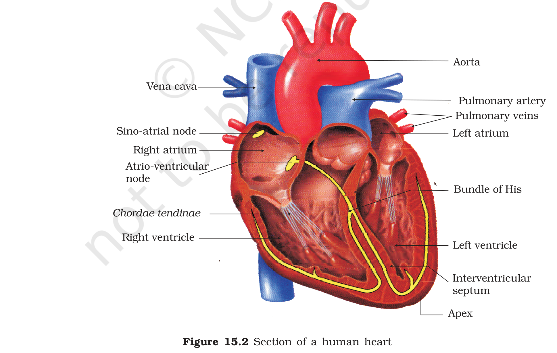
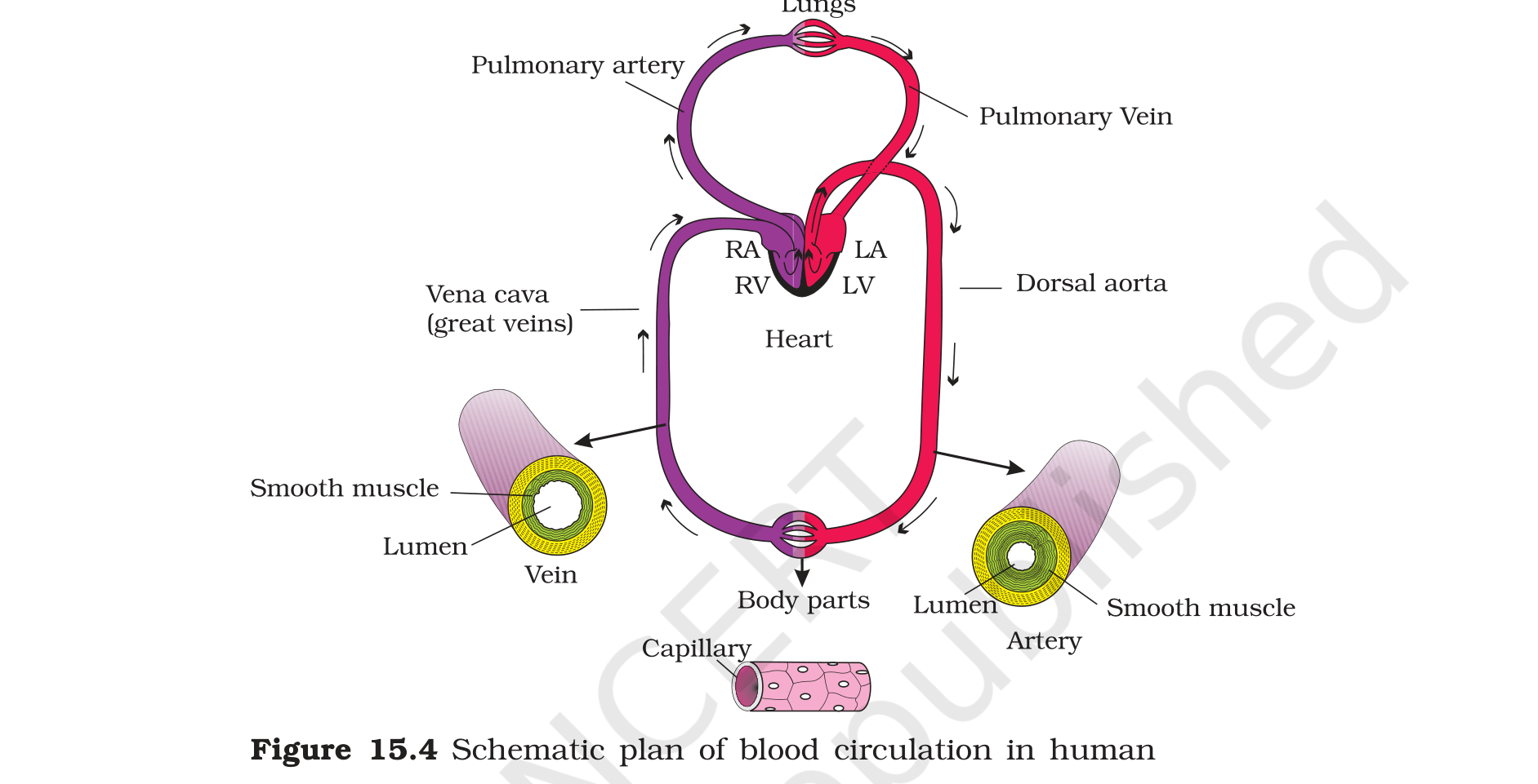
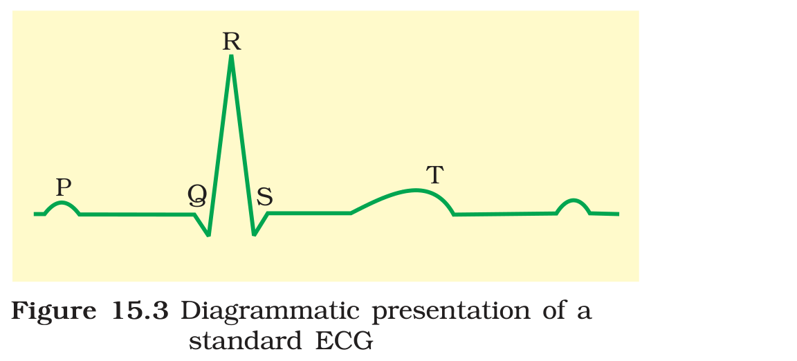
Figure 15.2: section of a human heart with the atria, ventricles, valves, sino-atrial node, atrio-ventricular node and bundle of His labelled
3.1.4 Listening to the cycle
The cardiac cycle can be followed at three levels at once. Mechanically, the chambers contract and relax in the order atria first and ventricles next. Electrically, the impulse passes from the sino-atrial node through the atria, then through the atrio-ventricular node, the bundle of His and the Purkinje fibres to the ventricles. Acoustically, the two valve closures give the two heart sounds. A problem in one level shows up in the others, which is why the examination combines a stethoscope, a pulse check and an ECG. A slow pulse with a normal ECG pattern suggests a conduction block, while an irregular ECG suggests an arrhythmia, and both findings change the wording of the final diagnosis.
3.1.5 Reading a cardiac report
A printed ECG report gives the paper speed, the rhythm and the duration of each wave, and the clinician compares these with the expected values. A normal beat starts with a P wave, follows with the QRS complex and ends with the T wave, and the intervals between the waves carry as much meaning as the waves themselves. A long PR interval suggests a slow passage of the impulse from the atria to the ventricles, while a widened QRS complex suggests a slower spread through the ventricular muscle. The same trace also shows whether the rhythm is regular, because the distance between successive R peaks is constant in a normal resting heart. Learning to read a few of these features turns the ECG from a picture into a piece of evidence.
Exam
A cardiac cycle answer should give the three phases with their durations and the events in each, then define stroke volume and cardiac output with the numbers. If the question asks for heart sounds, link the first sound to the closing of the atrioventricular valves and the second sound to the closing of the semilunar valves. For the ECG, write P, QRS and T in order with atrial depolarisation, ventricular depolarisation and ventricular repolarisation.
Common mistake
Do not say that the atria and ventricles contract together in a normal cycle, because atrial systole ends before ventricular systole begins. Do not place the pacemaker in the atrio-ventricular node; the sino-atrial node sets the rhythm and the AV node delays the impulse so that the atria can empty first. Do not say that the heart needs a nerve impulse to start each beat, because the heartbeat is myogenic; nerves only speed it up or slow it down.