

Concepts: The neural system, its divisions and the neuron
Neural Control and Coordination24 min readLesson 1 of 5
1.1 Coordination by the nervous system
An animal has to adjust its internal working to the outside world, and it does so through two coordinating systems: the neural system, which is fast and uses electrical signals, and the endocrine system, which is slower and uses hormones. The neural system is built of specialised cells called neurons, which are excitable: their membrane can change its electrical state in response to a stimulus and pass the change along. The neural system receives information from the sense organs, processes it in the brain and the spinal cord, and sends commands to the muscles and glands.
The human neural system is divided into the central neural system and the peripheral neural system. The brain and the spinal cord together make the central neural system, and information is processed and decisions are taken there. The nerves that link the central system with the rest of the body make up the peripheral neural system, which is split into two divisions. The somatic neural system carries impulses to the skeletal muscles, which are under conscious control, so it looks after voluntary movement. The autonomic neural system carries impulses to the smooth muscles, the heart and the glands, which work without our conscious control, and it is further divided into the sympathetic and the parasympathetic divisions. These two divisions have opposite effects on the same organs, so one speeds the heart and the other slows it, and the balance between them keeps the internal state steady.
1.2 Neurons and their supporting cells
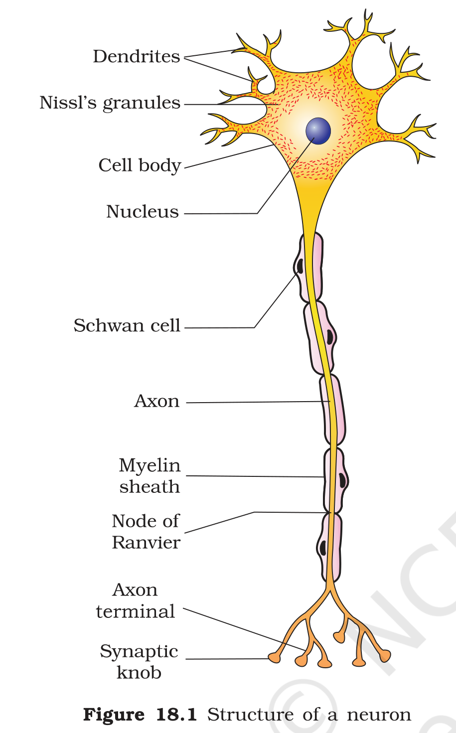
A neuron has three parts: the cell body, the dendrites and the axon. The cell body, also called the soma, contains the nucleus and the cytoplasm with ordinary cell organelles, and it also has certain granular bodies called Nissl's granules. Short branched processes, the dendrites, pick up signals from neighbouring neurons and pass them inwards to the cell body. One long process, the axon, then takes the impulse outwards towards the next cell in the chain. At its far end the axon branches into fine endings, and each ending holds a swollen synaptic knob which contains vesicles of neurotransmitter.
The axon of many neurons is covered by a myelin sheath. In the peripheral nervous system the sheath is formed by Schwann cells, which wrap themselves around the axon, and the sheath is interrupted at intervals by small gaps called the nodes of Ranvier. The myelin covering acts as an insulator, so the current leaks out of the axon very little, and in a myelinated axon the impulse can jump from one node to the next, which is much faster than travelling continuously along an uncovered membrane. The regions between the nodes carry the ion channels that allow the impulse to be regenerated.
Neurons are classified by the number of processes that leave the cell body. A multipolar neuron has one axon and several dendrites and is the commonest type in the brain and spinal cord. A bipolar neuron has one axon and one dendrite and is found in the retina of the eye. A unipolar neuron has a cell body with a single axon, as in the early embryo. The neurons work together with supporting cells called neuroglia, which protect and insulate them and keep their surroundings suitable for signalling.
1.2.1 Two coordinating systems
The neural system is not the only coordinator of the body. The endocrine system also controls and integrates the working of organs, but it does so with hormones carried in the blood. The two systems differ in speed and in reach. A nerve impulse travels in milliseconds and its effect is limited to the cell that the axon touches, while a hormone may take seconds to minutes to act and it can reach every cell that carries the matching receptor. The two systems also work together: the hypothalamus is a part of the brain, yet it controls the pituitary gland, so a neural signal can start a hormonal cascade. This is why the chapter on neural control and the chapter on chemical coordination describe two halves of one regulatory network rather than two unrelated systems.
1.2.2 Supporting cells around the neurons
Neurons do not work alone. They are surrounded by neuroglial cells, which are far more numerous than the neurons themselves. These cells hold the neurons in place, supply them with nutrients, remove their wastes, and in the case of the Schwann cells and their central counterparts they wrap the axons in myelin. Some glial cells also line the fluid filled spaces of the brain and help to regulate the composition of the fluid around the neurons. An axon that loses its myelin loses much of its conducting speed, which is why diseases that damage myelin produce weakness and loss of coordination.
Exam
For the divisions of the neural system, draw a tree: neural system splits into central (brain and spinal cord) and peripheral, and the peripheral splits into somatic, which goes to skeletal muscle, and autonomic, which goes to smooth muscle, heart and glands and is itself split into sympathetic and parasympathetic. For the neuron, give the job of each part: dendrite receives, axon conducts, synaptic knob releases the transmitter, myelin insulates and the node of Ranvier lets the impulse be regenerated.
Common mistake
Do not say that the somatic nervous system controls the internal organs, because it controls skeletal muscles and voluntary movement. Do not confuse the sympathetic and parasympathetic divisions, which have opposite effects on the same organ. Do not say that dendrites carry impulses away from the cell body, since they carry them towards it. Also do not call the myelin sheath a continuous covering, because it is interrupted at the nodes of Ranvier.