

Concepts: Anatomy of the leaf and exercise practice
Anatomy of Flowering Plants18 min readLesson 3 of 5
3.1 The leaf in section
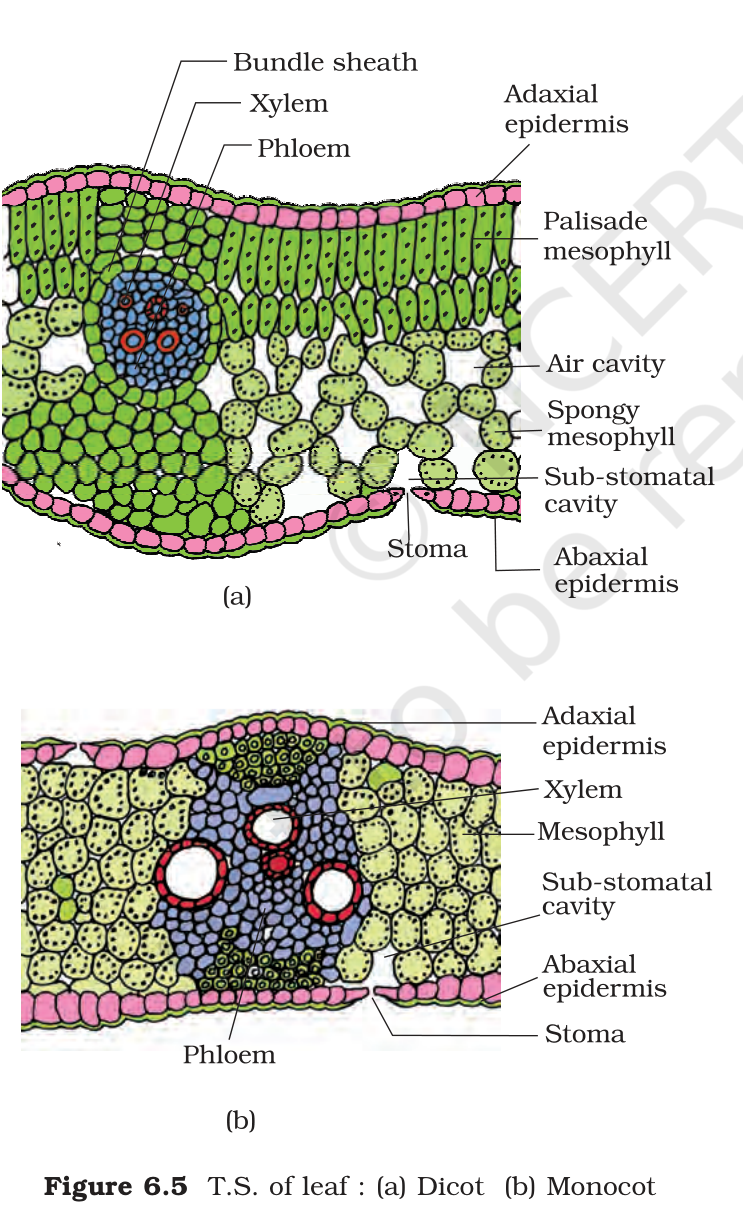
A leaf is read from the upper surface to the lower surface. Three parts make the section: the epidermis above and below, the mesophyll between them, and the vascular system embedded in the mesophyll. The pattern differs between a dorsiventral leaf, which is the usual dicot leaf, and an isobilateral leaf, which is the usual monocot leaf.
3.1.1 The dorsiventral leaf
The epidermis covers both surfaces. The upper surface is the adaxial side and the lower surface is the abaxial side, and both carry a clear cuticle. The lower epidermis usually has more stomata than the upper one, and in some plants the upper epidermis has none at all. Between the two epidermal layers lies the mesophyll, the ground tissue of the leaf, made of parenchyma with chloroplasts. The mesophyll has two cell types. The palisade parenchyma lies towards the upper surface, built of elongated cells arranged vertically and side by side. The spongy parenchyma lies below it, made of oval or rounded cells placed loosely with large air spaces between them, and it reaches down to the lower epidermis. Bundles run inside the veins and the midrib, each wrapped by a sheath of thick-walled cells. A bigger vein carries a bigger bundle, so a reticulate leaf shows bundles of many sizes.
3.1.2 The isobilateral leaf
The isobilateral leaf follows the same plan in many ways, and its differences are the ones to learn. Stomata are present on both surfaces. The mesophyll is not divided into palisade and spongy parts, so the tissue looks alike from either side, which is why the leaf is called isobilateral. In grasses a row of large, empty, colourless cells runs along the veins in the upper epidermis; these are the bulliform cells. When they are turgid with water the leaf surface stays open, and when they lose water they shrink and roll the leaf inward, cutting water loss. The parallel venation shows in the near-equal size of the vascular bundles, apart from the main veins.
3.1.3 Why anatomy is useful
Anatomy is more than a practical skill. It lets a person identify a plant organ from a bit of material, tell monocots from dicots in the field, and judge the quality of wood and fibres. It also explains how a plant works: the cuticle limits water loss, bulliform cells roll a grass leaf in a dry spell, and the air spaces of spongy tissue keep a leaf light and ventilated. When a question asks for the usefulness of anatomy, answer with these examples rather than with a general claim.
3.1.4 Practice from the NCERT exercises, questions 6 and 7
Exercise 6. The study of plant anatomy helps in several ways. It identifies plant material from small fragments, separates monocot from dicot organs, links structure to function such as cuticle to water loss and bulliform cells to leaf rolling, and helps in judging wood, fibres and other plant products.
Exercise 7. A labelled diagram of a dorsiventral leaf should show the upper epidermis with cuticle, the lower epidermis with cuticle and stomata, the palisade parenchyma below the upper epidermis, the spongy parenchyma below it with air spaces, and a vascular bundle with xylem and phloem wrapped in a bundle sheath.
3.1.5 The two leaf types compared
| Feature | Dorsiventral leaf | Isobilateral leaf |
|---|---|---|
| Stomata | mostly on the lower surface | on both surfaces |
| Mesophyll | palisade above, spongy below | not divided into two types |
| Bulliform cells | absent | present in the upper epidermis of grasses |
| Example | most dicots | most monocots |
The table is the shortest way to hold the two leaf plans. The dorsiventral leaf divides its work between a light-facing upper layer and a gas-exchanging lower layer. The isobilateral leaf gives both surfaces the same structure, and in grasses it adds bulliform cells that let the blade fold when water is short. The veins differ too: a dicot leaf shows veins of many sizes, while a monocot leaf shows many similar parallel bundles. On a slide the bundle sheath around each vein is the easiest landmark, because it stands out as a ring of thick-walled cells wherever a vein passes.
3.1.6 The stem sections in one table
| Feature | Dicot stem | Monocot stem |
|---|---|---|
| Hypodermis | collenchymatous | sclerenchymatous |
| Vascular bundles | in a ring, conjoint and open | scattered, conjoint and closed |
| Bundle sheath | absent | sclerenchymatous sheath present |
| Medullary rays | present | absent |
Two characters settle most practical questions: the ring of bundles points to a dicot stem and the scattered bundles point to a monocot stem, while the bundle sheath confirms the second. The hypodermis and the medullary rays are the supporting evidence.
Exam
For the leaf section, the marks sit on three differences between a dorsiventral and an isobilateral leaf: stomata on one surface versus both, mesophyll divided into palisade and spongy versus not divided, and bulliform cells present in grasses. Draw both sections side by side with the labels, because the diagram itself carries marks in this question.
Common mistake
Do not write that both leaves have palisade tissue, because the monocot leaf does not divide its mesophyll that way. Do not put the bulliform cells in the lower epidermis, since they lie on the upper surface along the veins of grasses. And do not say the vascular bundle lies above the epidermis, because it sits inside the mesophyll.