

Concepts: Concentration of urine and hormonal regulation
Excretory Products and their Elimination24 min readLesson 3 of 6
3.1 How the kidney concentrates urine
Filtered fluid leaves the loop of Henle diluted, and the kidney has to draw water out of it before the urine is passed. It manages this by keeping the tissue of the inner medulla unusually salty, and the arrangement that builds and protects that salt is the counter current mechanism.
Two counter currents work together. In the loop of Henle the fluid runs down one limb and up the other, so the two flows point in opposite directions. In the vasa recta, the small vessels that follow the loop, the blood also runs in opposite directions in its two limbs. Because the loop and the vasa recta lie side by side, the salt that leaves the loop can be caught by the blood and returned to the medulla instead of being carried away.
The result is a saltiness that climbs from about 300 mOsmol per litre in the cortex to about 1200 mOsmol per litre deep in the medulla. Two substances hold the gradient in place. Sodium chloride is moved out of the ascending limb of the loop and swapped with the descending limb of the vasa recta, and the ascending part of the vasa recta puts the salt back into the medullary tissue. Urea follows a smaller loop of its own: a little of it enters the thin part of the ascending limb, and the collecting tubule carries it back into the interstitium. The water that leaves the collecting duct is carried away by the vasa recta without flushing the salt and urea out of the medulla. Since the surrounding tissue is hypertonic, water leaves the collecting duct readily and the urine becomes concentrated. In a human kidney the final urine can be about four times as concentrated as the filtrate that first entered the tubule.
3.2 Hormones that regulate the kidney
Three hormonal loops adjust the working of the kidney, and each loop has its own trigger and its own effect.
3.2.1 Water and antidiuretic hormone
The hypothalamus contains osmoreceptors that notice changes in the volume of the body fluids and in their ionic concentration. When the body loses a lot of fluid, these receptors fire and the hypothalamus instructs the neurohypophysis to release antidiuretic hormone, also called vasopressin. The hormone makes the later parts of the tubule take back more water, which prevents diuresis. When the body fluid volume is restored, the receptors stop firing and the hormone release falls, so the loop closes. ADH has a second effect on the vessels: it narrows them, and the resulting rise in pressure can push the glomerular blood flow and the GFR upward.
3.2.2 Salt, pressure and the renin chain
The juxtaglomerular apparatus watches the blood pressure in the glomerulus. If the pressure, the flow or the GFR drops, the JG cells put out renin. Renin acts on angiotensinogen in the blood and starts a short chain that produces angiotensin I and then angiotensin II. Angiotensin II narrows the vessels strongly, and the narrowed vessels raise the pressure inside the glomerulus, which pulls the GFR back up. The same angiotensin II tells the adrenal cortex to release aldosterone, and aldosterone makes the distal tubule take back more sodium and water. The extra salt and water raise the blood pressure and the GFR still further. This chain is the renin-angiotensin mechanism.
3.2.3 The heart's brake
When more blood reaches the atria of the heart, the atria release atrial natriuretic factor, or ANF. ANF widens the blood vessels, and the wider vessels bring the pressure down, so ANF works against the renin chain and keeps it from running away. The three loops together keep the GFR, the blood pressure and the salt and water balance inside a range the body can live with.
3.2.4 Practice from the NCERT exercises, questions 4, 10 and 12
Exercise 4. The concentration depends on the loop of Henle and the vasa recta lying close together with their flows opposed. Salt is moved out of the ascending limb into the medullary tissue, the descending limb lets water pass, and the vasa recta drain the water away while the salt and urea stay behind. This builds the rising osmolarity from the cortex to the inner medulla, and the collecting duct can then pull water out of the filtrate so that the urine becomes concentrated.
Exercise 10. The juxtaglomerular apparatus is a sensitive region where the distal convoluted tubule touches the afferent arteriole. It senses the glomerular blood flow and pressure and releases renin when the GFR falls, and the renin-angiotensin-aldosterone mechanism then restores the pressure and the filtration rate.
Exercise 12. In the loop of Henle, the ascending limb does not let water through, while the descending limb does. The hormone that helps the later tubule take back water is antidiuretic hormone. The dialysing fluid is like plasma but without the nitrogenous wastes such as urea. A healthy adult passes roughly 25 to 30 g of urea in a day.
Slideshow, slide 1 of 3. Use the left and right arrow keys to change slides and the space bar to play or pause.
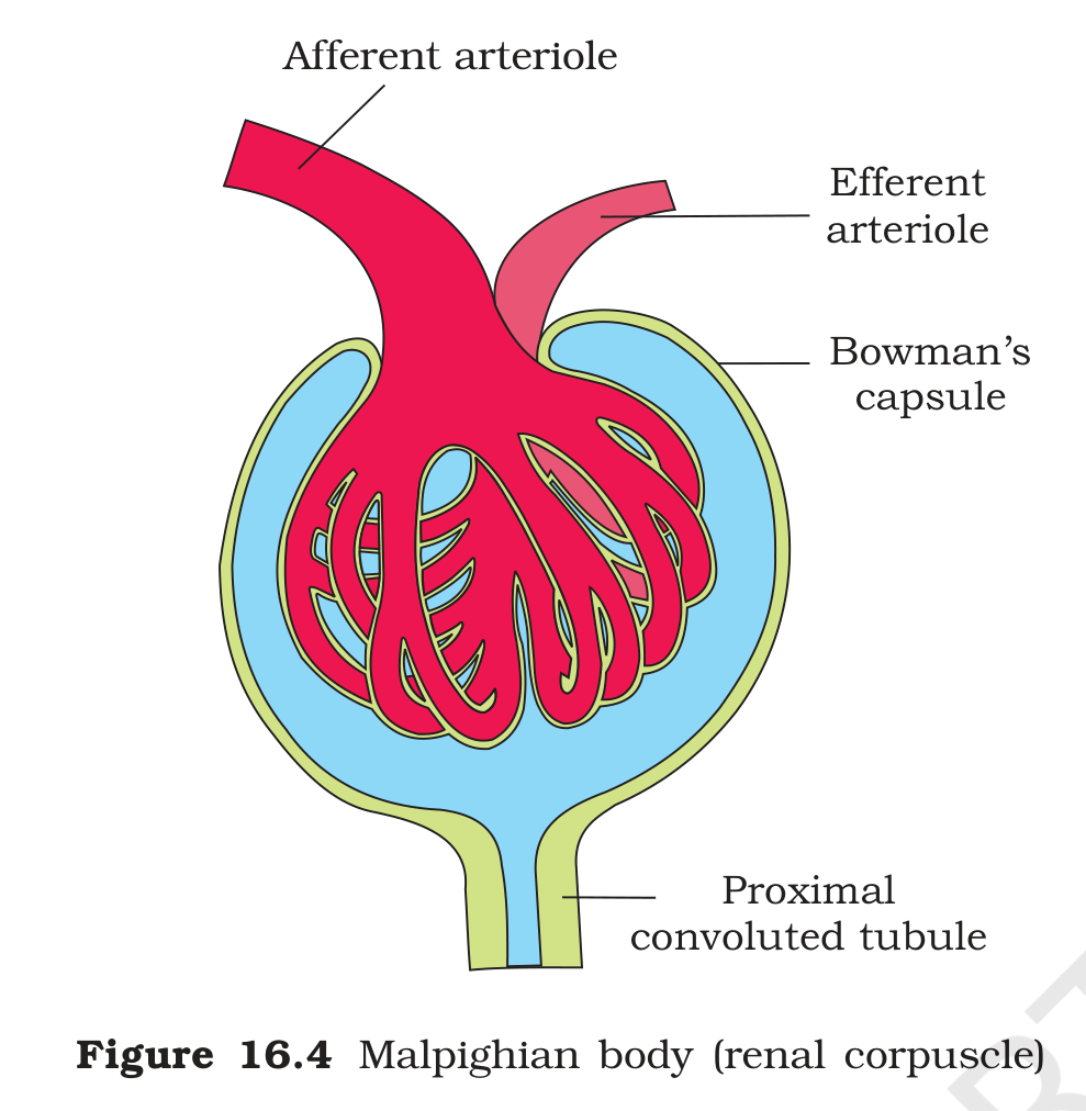
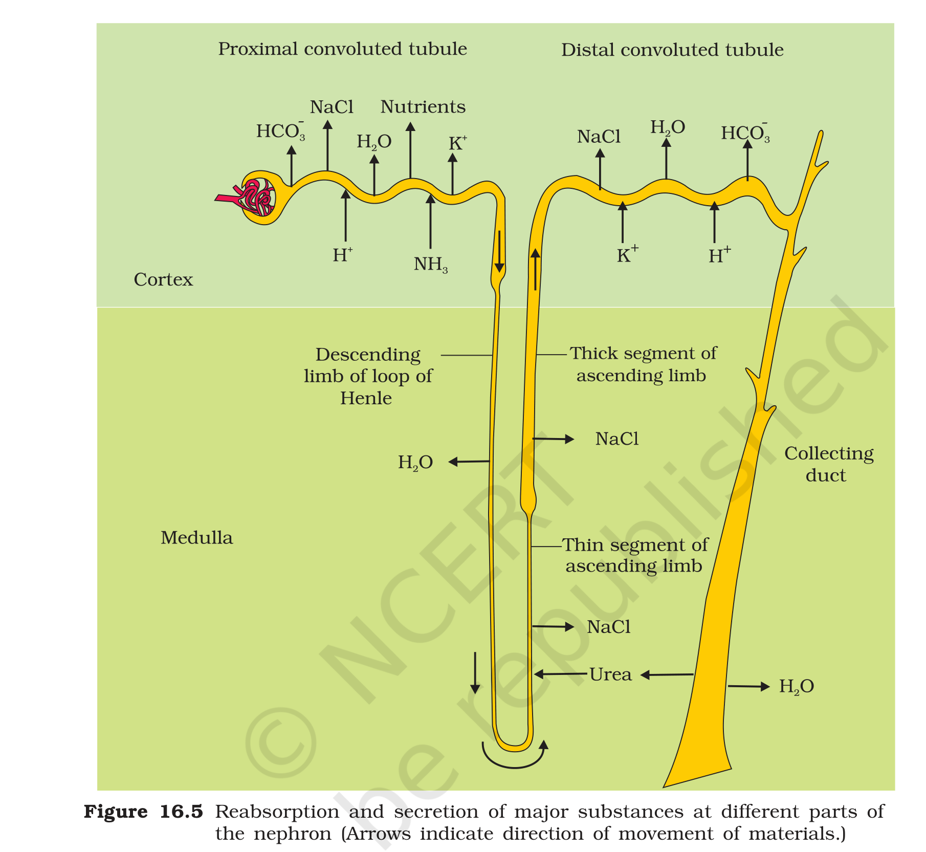
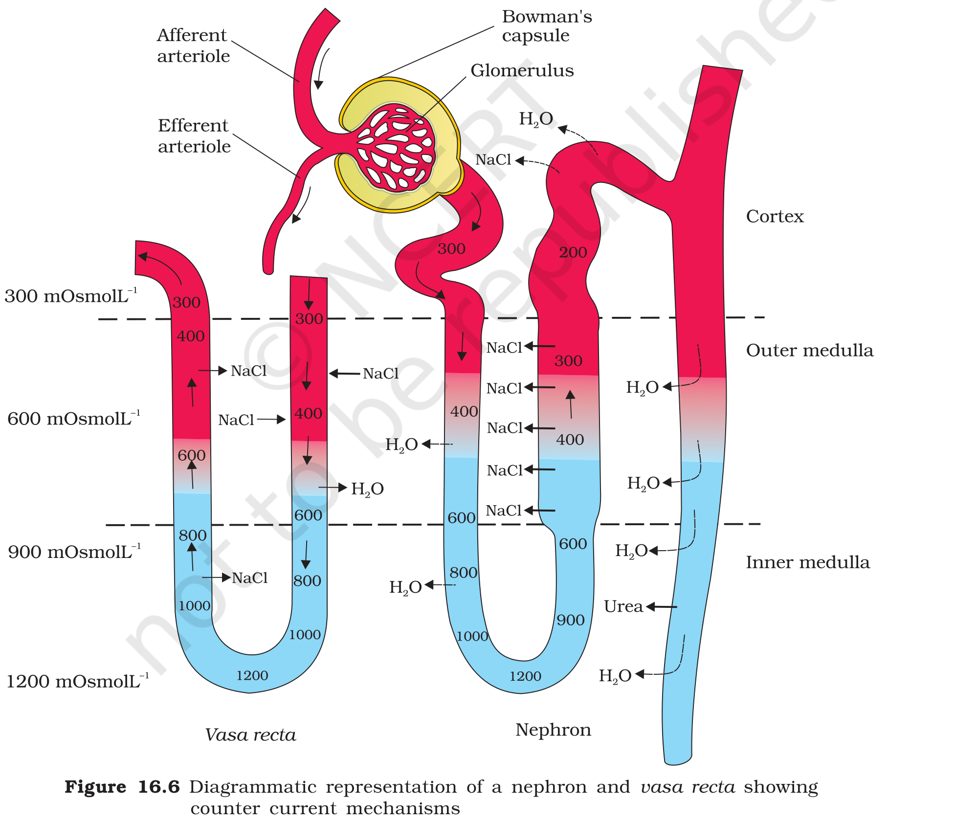
Figure 16.4: malpighian body or renal corpuscle with the afferent arteriole, efferent arteriole, Bowman's capsule, glomerulus and proximal convoluted tubule
Exam
For the counter current answer, describe both counter currents, then give the two numbers 300 and 1200 mOsmol per litre, then name the two solutes that maintain the gradient. Close with the sentence that the hypertonic medulla lets the collecting duct reabsorb water and concentrate the urine. For the hormonal answer, draw the three loops separately: ADH for water, renin-angiotensin-aldosterone for sodium and pressure, and ANF as the brake on the renin loop.
Common mistake
Do not say that the ascending limb is permeable to water. It is impermeable to water and moves salt out, which is exactly why it dilutes the filtrate. Do not say that aldosterone reabsorbs water directly; it acts mainly on sodium reabsorption, and water follows the sodium. Do not call ANF a hormone that raises blood pressure, because it lowers the pressure by causing vasodilation.