

Concepts: Respiratory organs and the human respiratory system
Breathing and Exchange of Gases22 min readLesson 1 of 6
1.1 Why breathing has to happen
Every cell needs oxygen for the oxidation of food and gives out carbon dioxide as a waste product. In a single-celled organism the two gases simply diffuse across the cell surface. In a large, multicellular animal the distance from the surface to the internal cells is too great for diffusion alone, so a specialised system moves air close to the blood and the blood carries the gases to and from the tissues. Breathing, or pulmonary ventilation, is the first step in this chain. The full process of respiration in the body has several steps: pulmonary ventilation moves air in and out of the lungs, diffusion across the alveolar membrane exchanges oxygen and carbon dioxide between air and blood, the blood transports the gases, diffusion between blood and tissue exchanges them again, and the cells finally use oxygen in catabolic reactions and release carbon dioxide.
1.1.1 Respiratory organs across animals
The organ used for gas exchange depends on the body plan and the habitat. Earthworms use the moist skin, and the skin must stay moist for the gases to dissolve and diffuse. Insects use a system of air-filled tubes, the tracheae, which open on the body surface through small openings called spiracles and branch until they reach the tissues, so their respiration is called direct. Fishes use gills, where water flows over the gill filaments and the gases diffuse between water and blood. Frogs use the moist skin, the lining of the mouth and the lungs. Birds and reptiles have lungs. These examples are the standard matching question for this chapter.
1.1.2 The human respiratory system
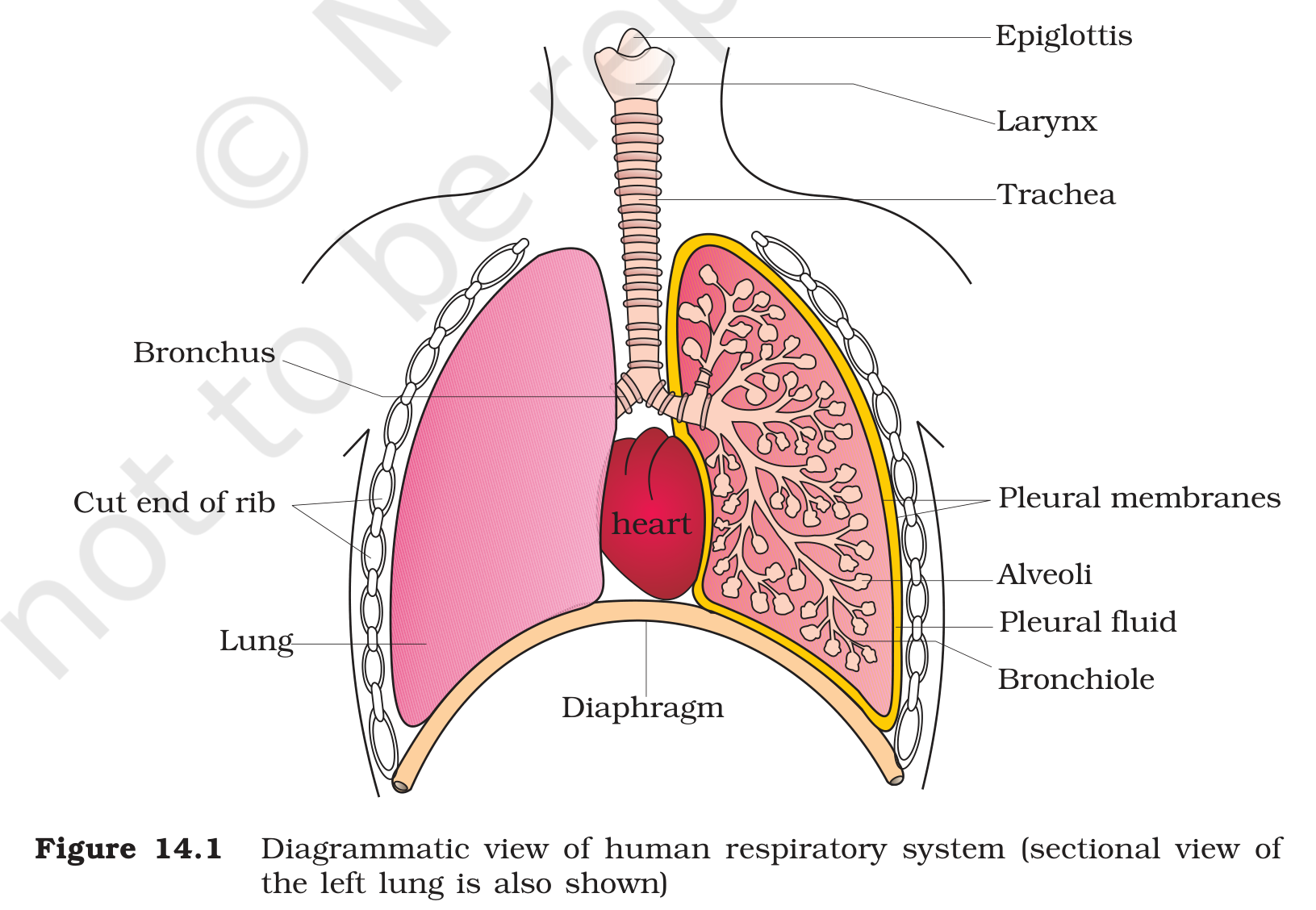
Air enters through the nostrils into the nasal cavity, where it is warmed, moistened and cleaned by the mucus and hairs. It then passes the pharynx, the larynx or voice box, the trachea, and the two primary bronchi that enter the lungs. Inside each lung the bronchi divide repeatedly into secondary and tertiary bronchi and finally into fine bronchioles, which end in clusters of thin-walled air sacs called alveoli. The alveoli are the actual sites of gas exchange, and their walls are one cell thick and closely wrapped by a network of blood capillaries. The lungs are covered by a double membrane, the pleura. The space between the two pleural layers holds pleural fluid, which reduces friction as the lungs move during breathing. The rib cage protects the lungs, and the diaphragm, a dome-shaped muscle, forms the floor of the chest cavity. Figure 14.1 shows the whole arrangement in section, with the bronchus, alveoli, pleural membranes, diaphragm and the cut end of a rib labelled. The exchange surface must be large in relation to the body size, thin enough for quick diffusion, moist enough to keep the gases dissolved and well supplied with blood. The alveolar region meets all four conditions, which is why diffusion of gases happens there and not in the nose or the trachea.
1.1.3 How the airways protect the lungs
The nose does more than admit air. Hairs at the entrance and the sticky mucus lining the nasal cavity trap dust particles, and the blood vessels in the lining warm and moisten the incoming air so that the alveoli receive a gentle, humid stream rather than cold dry air. The trachea is a tube that carries the air down to the middle of the chest, where it splits into the two primary bronchi, one for each lung. The branching bronchi and bronchioles form a tree that spreads through the lung tissue and ends in the alveolar clusters.
1.1.4 What makes an exchange surface efficient
Four features make the alveolar region suitable for exchange. The surface area is very large because there are millions of alveoli, so plenty of membrane is available for diffusion. The barrier is thin, only one cell of epithelium plus the capillary endothelium, which keeps the diffusion path short. The surface is moist, so the gases dissolve before crossing. Finally, the capillary network keeps blood flowing past the alveoli so that the concentration gradient does not collapse as the blood loads oxygen. If any one of these features fails, exchange slows, which is exactly what happens in diseases that damage the alveolar wall.
1.1.5 Ventilation keeps the gradient alive
Diffusion would stop if the air inside the alveoli came to the same composition as the blood. Breathing prevents that by replacing the alveolar air again and again. Each inspiration brings fresh air with a high pO2 and a low pCO2, and each expiration carries away the air that has already picked up carbon dioxide. The continuous flow of blood through the alveolar capillaries removes the oxygen as it crosses, which keeps the pO2 in the blood below the pO2 in the air. Ventilation and blood flow therefore keep both gradients steep, and the rate of exchange depends on how well these two flows are matched.
Exam
For the matching question on respiratory organs, write earthworm with moist skin, insects with tracheae and spiracles, fishes with gills, and frogs with skin, mouth lining and lungs. For the human system, the order is nostrils, nasal cavity, pharynx, larynx, trachea, bronchi, bronchioles and alveoli, and the exchange happens only in the alveoli.
Common mistake
Do not say that gas exchange happens all along the respiratory tract. The nose, trachea and bronchi only warm, moisten, clean and conduct the air; the thin, blood-rich alveolar surface is the exchange region. Do not confuse the pleura with the peritoneum, and do not place the diaphragm above the lungs. Insects do not use lungs, and their tracheae carry air directly to the tissues.