

Concepts: Gametogenesis and the menstrual cycle
Human Reproduction24 min readLesson 3 of 8
3.1 Spermatogenesis
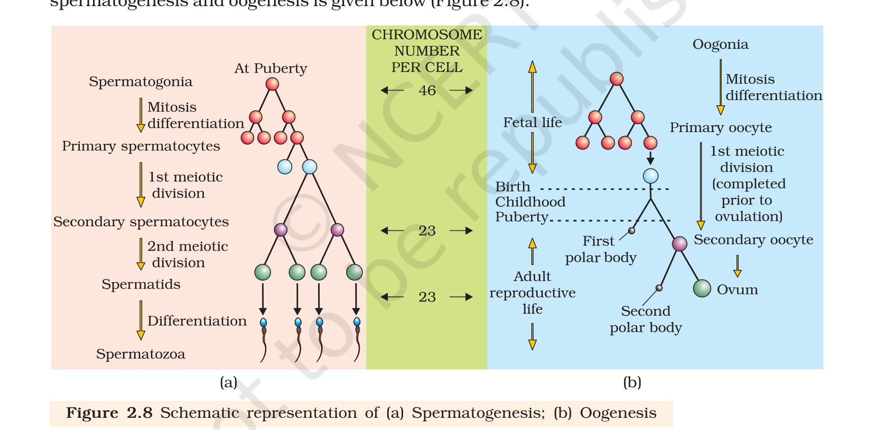
Gametogenesis is the formation of gametes by the primary sex organs, the testes in the male and the ovaries in the female. In the testis the immature germ cells are the spermatogonia, and spermatogenesis begins there at puberty. Spermatogonia lie on the inner wall of the seminiferous tubules, divide by mitosis and increase in number. Each spermatogonium is diploid and carries 46 chromosomes.
Some spermatogonia, now called primary spermatocytes, periodically enter meiosis. The first meiotic division is a reduction division and gives two equal haploid cells, the secondary spermatocytes, each with 23 chromosomes. The second meiotic division then produces four equal haploid spermatids from each primary spermatocyte. The spermatids are remodelled into spermatozoa, and that transformation is called spermiogenesis. After spermiogenesis the sperm heads sit embedded in the Sertoli cells, and the mature sperms are finally released from the tubule by spermiation.
3.2 Hormones that switch on sperm production
Spermatogenesis starts at puberty because the hypothalamus begins to secrete more gonadotropin releasing hormone, or GnRH. It prompts the anterior pituitary to release more of the two gonadotropins: luteinising hormone, or LH, and follicle stimulating hormone, or FSH. LH targets the Leydig cells and pushes them to make and release androgens. The androgens push spermatogenesis along. FSH targets the Sertoli cells and induces them to secrete factors that assist spermiogenesis. The chain is short and worth writing exactly: GnRH, then LH and FSH, then Leydig cells and androgens, then sperm.
3.3 The sperm
A sperm is tiny and divided into four parts: head, neck, middle piece and tail, with the plasma membrane wrapping the whole cell. The head carries an elongated haploid nucleus, and the front of the nucleus is capped by the acrosome, a structure filled with enzymes that digest a path into the ovum during fertilisation. The middle piece is packed with mitochondria, and these supply the energy for the tail to beat. The tail gives the sperm its motility, which is essential for fertilisation.
The numbers here are worth remembering. A human male ejaculates about 200 to 300 million sperms in one copulation. For normal fertility, at least six out of every ten must look normal in shape and size, and at least four out of every ten must swim actively. Secretions from the epididymis, vas deferens, seminal vesicle and prostate allow the sperm to mature and keep moving.
Slideshow, slide 1 of 3. Use the left and right arrow keys to change slides and the space bar to play or pause.
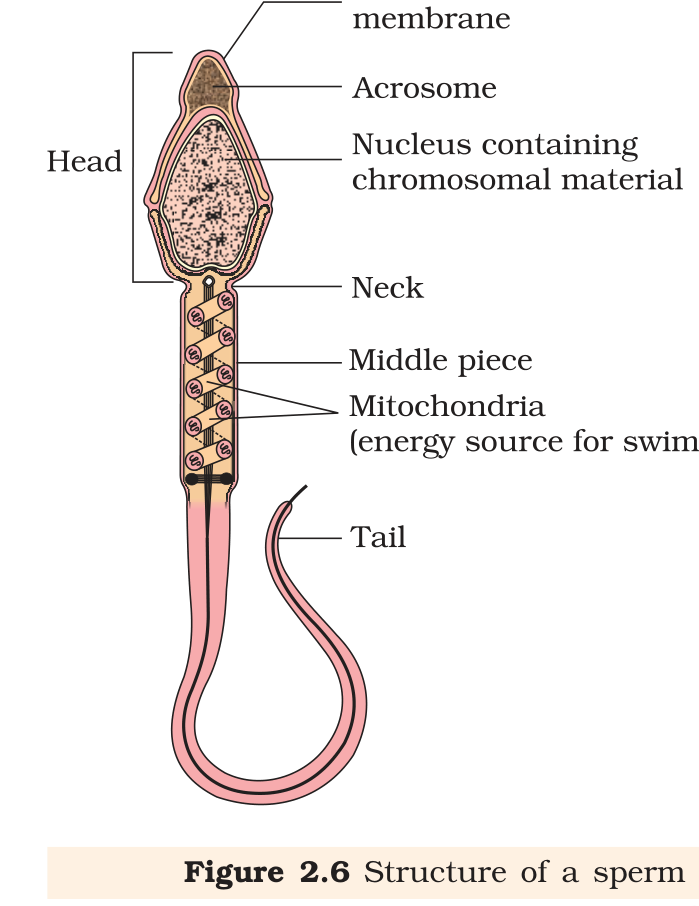
Structure of a sperm with the head, acrosome, nucleus, neck, middle piece and tail labelled.
3.4 Oogenesis
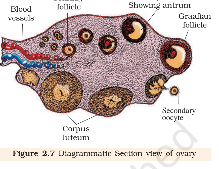
Oogenesis is the formation of the mature female gamete, and it differs from spermatogenesis in several important ways. It begins during embryonic development, when a few million oogonia form in each fetal ovary. No new oogonia are added after birth. These cells start dividing, enter prophase I of meiosis and stop there. A cell arrested at this stage is a primary oocyte. A single layer of granulosa cells then wraps the primary oocyte, and the unit becomes a primary follicle.
A large number of follicles degenerate between birth and puberty, and only about 60,000 to 80,000 primary follicles remain in each ovary at puberty. More granulosa layers and a fresh theca turn the primary follicles into secondary ones. A secondary follicle soon becomes a tertiary one, recognisable by a fluid-filled space, the antrum. By now the theca has split into an inner interna and an outer externa.
The primary oocyte inside the tertiary follicle finally completes its first meiotic division. This division is unequal. It produces one large haploid secondary oocyte, which keeps almost all of the nutrient-rich cytoplasm, and one tiny first polar body. The secondary oocyte then forms a new membrane around itself called the zona pellucida, and the tertiary follicle becomes the mature Graafian follicle. When the Graafian follicle ruptures, the secondary oocyte is released from the ovary, and that release is ovulation.
| Feature | Spermatogenesis | Oogenesis |
|---|---|---|
| Starts | At puberty | Before birth, in the fetus |
| Products per meiosis | Four spermatids | One secondary oocyte and polar bodies |
| Cytoplasm in the products | Shared almost equally | Kept almost entirely by the oocyte |
| Completion | Continuous after puberty | Completed in stages, second division only after fertilisation |
| Release | Sperm released continuously | One ovum released per cycle |
3.5 The menstrual cycle
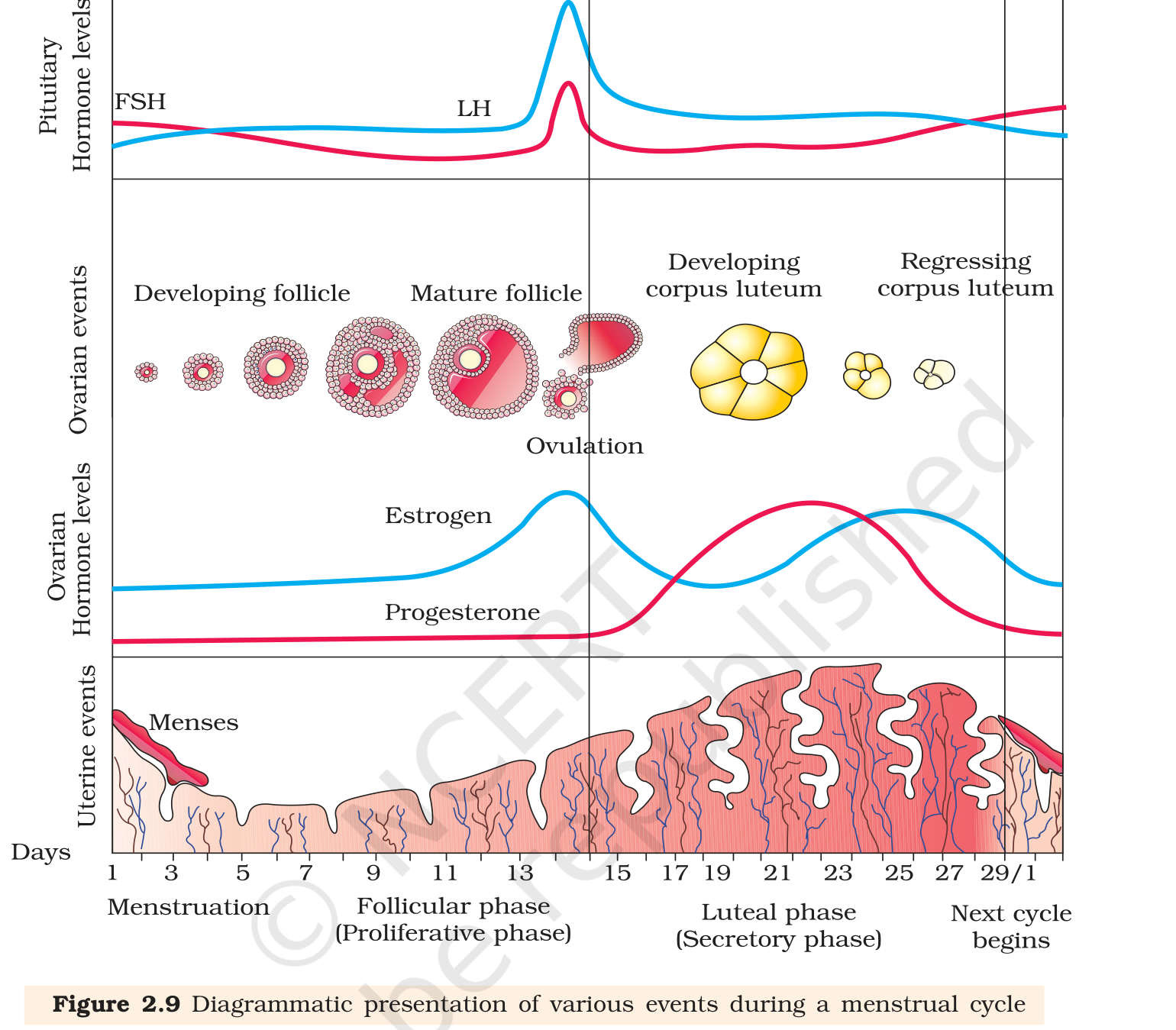
The reproductive cycle of female primates, including humans, is the menstrual cycle. The first menstruation, at puberty, is called menarche. In human females menstruation repeats at an average interval of about 28 or 29 days, and the cycle runs from one menstruation to the next. One ovum is released around the middle of the cycle.
The cycle begins with the menstrual phase, which lasts about three to five days. Menstrual flow happens because the lining of the uterus, the endometrium, and its blood vessels break down and the blood comes out through the vagina. This phase occurs only when the released ovum was not fertilised. A missed period may point to pregnancy, but stress and poor health can also delay it.
The follicular phase follows. Across this phase the ovarian primary follicles enlarge until one is a mature Graafian follicle, and the endometrium rebuilds itself by proliferation. The pituitary hormones LH and FSH rise gradually and stimulate follicular development and the secretion of oestrogens by the growing follicles. Both reach a peak in the middle of the cycle, around day 14.
The mid-cycle surge of LH is what triggers ovulation. The surge makes the Graafian follicle rupture and releases the ovum. That is the ovulatory phase.
The luteal phase follows ovulation. The remains of the Graafian follicle turn into the corpus luteum, which secretes a large amount of progesterone. Progesterone maintains the endometrium in the state needed for implantation of a fertilised ovum and for the early events of pregnancy. If fertilisation does not occur, the corpus luteum degenerates, the endometrium breaks down and menstruation begins again. If pregnancy does occur, the cycle stops and there is no menstruation. Menstrual cycles cease altogether around the age of fifty, and that stage is called menopause. Cyclic menstruation between menarche and menopause is a sign that the reproductive phase is normal.
Exam
For a five-mark question on the menstrual cycle, draw the four phases on a time line and put the hormone beside each phase: oestrogen in the follicular phase, an LH peak at ovulation, progesterone from the corpus luteum in the luteal phase. Name the two pituitary hormones and the two ovarian hormones and you have most of the marks.
Common mistake
Do not write that the oocyte divides equally. The first meiotic division is unequal, giving one large secondary oocyte and one small polar body. Also do not say that polar bodies are always wasted; the chapter notes that their later fate is not settled. And remember that the LH surge, not FSH alone, causes ovulation.