

Concepts: The female reproductive system and the mammary glands
Human Reproduction17 min readLesson 2 of 8
2.1 The female system as a whole
The female tract is built from an ovary on each side, an oviduct on each side, one uterus, a cervix, a vagina and the external genitalia, all within the pelvis. A pair of mammary glands completes the set. These organs act together for ovulation, fertilisation, pregnancy, birth and the care of the child, and that is why they are described as one integrated system.
The ovaries are the primary female sex organs. They make the ovum and also produce steroid hormones, the ovarian hormones. Each ovary lies on one side of the lower abdomen, is about two to four centimetres long, and is held to the pelvic wall and the uterus by ligaments. A thin epithelium covers the ovary and encloses the ovarian stroma. Two zones make up the stroma: a cortex at the rim and a medulla inside. Follicles at different stages of growth sit embedded in it.
The ovary carries the same double role as the testis. It produces the female gamete, the ovum, and it releases steroid hormones that act on the uterus and drive the cycle. Both jobs happen in the follicles of the cortex. Each follicle holds an oocyte, and the growing follicle releases oestrogen while the corpus luteum that forms after ovulation releases progesterone. An answer about ovarian function should therefore name the gamete and the hormone, and it should name the follicle and the corpus luteum as the structures involved.
2.2 Oviducts, uterus and vagina
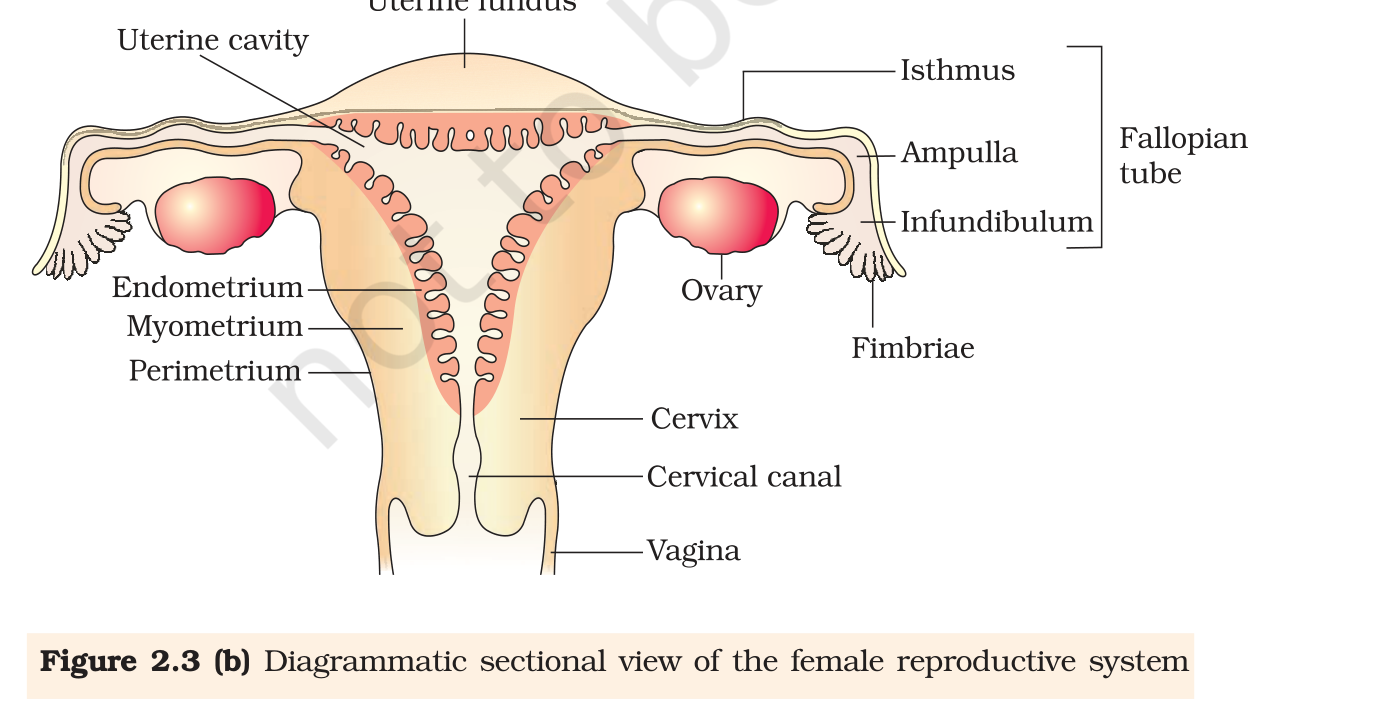
Each fallopian tube is about ten to twelve centimetres long and runs from the region of the ovary to the uterus. The end nearest the ovary is the funnel-shaped infundibulum, and its edges carry finger-like projections called fimbriae. The fimbriae sweep over the ovary and collect the ovum after ovulation. Past the infundibulum the tube widens into the ampulla, and its last part is a narrow isthmus that joins the uterus. Fertilisation normally happens in the ampulla.
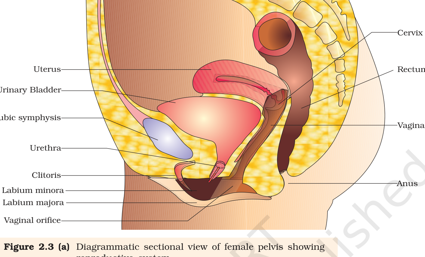
The uterus is single and is also called the womb. It is shaped like an inverted pear and is held by ligaments attached to the pelvic wall. It opens into the vagina through a narrow cervix, and the cavity of the cervix is the cervical canal. The cervical canal plus the vagina make up the birth canal.
Three tissue layers build the uterine wall. Outside is the thin membranous perimetrium. The middle myometrium is a thick layer of smooth muscle, and it contracts strongly during delivery. The inner endometrium is glandular and lines the uterine cavity, and it changes cyclically during the menstrual cycle. Two layers with two very different jobs, and an exam often asks which is which.
2.3 External genitalia
Outside, the genital structures are the mons pubis, the labia majora and minora, the hymen and the clitoris. Fat under skin, with pubic hair over it, forms the mons pubis. From the mons pubis two fleshy folds, the labia majora, run down and frame the vaginal opening. Under those lie the smaller paired folds, the labia minora. A membrane, the hymen, often covers part of the vaginal opening. Where the two labia minora meet at the top, above the urethral opening, sits a small finger-like body, the clitoris.
The hymen deserves a careful sentence, because biology and social belief often disagree here. It may tear during first intercourse, but it can also tear from a fall or a jolt, from inserting a vaginal tampon or from sports such as horse riding and cycling. In some women it stays intact even after intercourse. Its presence or absence is therefore not a reliable indicator of virginity or of sexual experience, and a board answer should say so plainly. Name the structures from the outside inward and the answer is complete.
2.4 Mammary glands
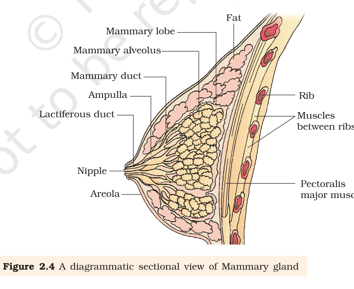
Every female mammal has functional mammary glands. In humans two breasts form a pair, holding glandular tissue mixed with fat in variable amounts. Each breast is divided into 15 to 20 mammary lobes, and each lobe carries clusters of milk-secreting cells called alveoli. The cells of the alveoli secrete milk into the cavities, or lumens, of the alveoli. Milk then leaves the alveoli for the mammary tubules, and within a lobe those tubules merge into one mammary duct. Several such ducts enlarge into a mammary ampulla, and from there a lactiferous duct opens at the nipple. Milk is expressed through the nipple during breastfeeding. The alveoli are the working units of the gland, and the ducts are the route that carries their secretion to the nipple. Each lobe drains through its own duct, so the gland works as a set of 15 to 20 drainage units that share one exit.
Exam
The three layers of the uterine wall are a favourite one-mark question. Remember the order from outside in: perimetrium, myometrium, endometrium. Give the function with each name, because the examiner looks for the link between the layer and either delivery or the menstrual cycle.
Common mistake
Do not state that the hymen proves virginity. The chapter says the opposite. Also keep the oviduct parts in order, infundibulum, ampulla and isthmus, and remember that fertilisation happens in the ampulla, not in the uterus.