

Concepts: The male reproductive system
Human Reproduction18 min readLesson 1 of 8
1.1 The sequence of human reproduction
Humans reproduce sexually and the young develop inside the mother, which makes humans viviparous. The events run in a fixed order. Gametogenesis makes the gametes, sperms in the male and ova in the female. Insemination delivers semen into the female genital tract. Fertilisation fuses the two gametes into a zygote. The zygote becomes a blastocyst, which attaches to the uterine wall, and that attachment is implantation. The embryo then develops through gestation, and the baby is delivered at parturition.
One difference between the sexes appears early in any discussion. Sperm formation begins at puberty and continues into old age, while a woman's store of ova is laid down before birth and her capacity to release them ends around the age of fifty. That asymmetry explains why the male and the female systems need separate descriptions.
1.2 Testes and the scrotum
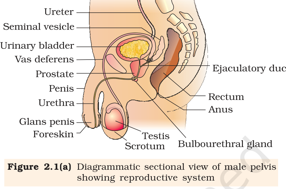
The male reproductive system lies in the pelvic region and consists of a pair of testes, the ducts that carry sperm, the accessory glands and the external genitalia. The testes sit outside the abdominal cavity in a pouch called the scrotum. The position matters: the scrotum keeps the testes about two to two and a half degrees below normal body temperature, and that lower temperature is needed for spermatogenesis. A testis that stays inside the abdomen cannot make fertile sperm.
Each adult testis is oval, about four to five centimetres long and two to three centimetres wide, and it has a dense fibrous covering. Inside, the testis is divided into roughly 250 compartments called testicular lobules. Each lobule holds one to three highly coiled seminiferous tubules, and sperm are produced inside these coils.
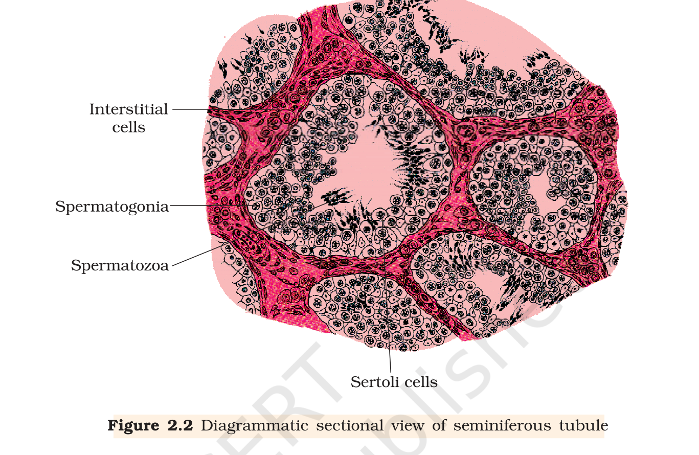
The wall of a seminiferous tubule is lined by two kinds of cells. The male germ cells, called spermatogonia, multiply by mitosis, and their descendants enter meiosis and eventually become sperm. The Sertoli cells sit among the germ cells and feed them as they develop. Outside the tubules are the interstitial spaces, packed with small blood vessels and interstitial cells, also called Leydig cells. Leydig cells make androgens, the testicular hormones. Immunologically competent cells are present in the same spaces.
The testis does two jobs, and a good answer keeps them apart. It produces the male gamete, the sperm, inside its seminiferous tubules. It also works as an endocrine gland, because the Leydig cells between the tubules make androgens. The Sertoli cells belong to the first job: their main role is support and nutrition, not androgen production. A question that asks for the functions of the testis usually wants both the gamete and the hormone, with the cell type named for each.
1.3 The duct system
Sperm made in the seminiferous tubules leave the testis through a pathway worth learning in order. The tubules open into the rete testis, which drains into the vasa efferentia. The short ducts feed a long coiled tube, the epididymis, that hugs the back of each testis. Beyond it the tube becomes the vas deferens, which rises into the abdomen and arches across the top of the urinary bladder. A branch from the seminal vesicle joins it there, and the joint tube enters the urethra as the ejaculatory duct. The urethra itself starts at the urinary bladder, travels the length of the penis and ends at the urethral meatus.
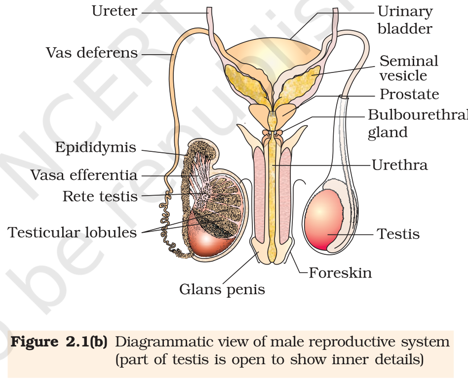
These ducts do more than carry sperm. They store the sperms and transport them out of the body, and their secretions, together with those of the accessory glands, keep the sperm alive and motile. Sperm that spend time in the epididymis mature and become motile there, which is why the duct is not a simple pipe.
1.4 Penis and accessory glands
The penis is the male external genitalia. It contains special erectile tissue that fills with blood and makes the penis erect, which is needed for insemination. The widened tip is the glans penis, and a loose skin fold, the prepuce or foreskin, covers it.
The accessory glands are the paired seminal vesicles, a single prostate and paired bulbourethral glands. Their secretions form the seminal plasma, a fluid rich in fructose, calcium and several enzymes. Fructose supplies energy to the sperm, calcium supports their activity and the enzymes help the semen to clot and later liquefy. The bulbourethral secretions also lubricate the penis. The seminal plasma together with the sperms makes up semen. On a diagram, the prostate sits below the urinary bladder and surrounds the first part of the urethra, and the bulbourethral glands lie just below the prostate.
| Part | Main role |
|---|---|
| Scrotum | Keeps the testes 2 to 2.5 degrees below body temperature |
| Seminiferous tubule | Site of spermatogenesis |
| Sertoli cells | Nutrition and support for developing germ cells |
| Leydig cells | Synthesise and secrete androgens |
| Epididymis | Sperm maturation and storage |
| Seminal vesicles, prostate, bulbourethral glands | Secretions that form seminal plasma |
Exam
A common three-mark question asks for the functions of the scrotum, the Leydig cells and the male accessory glands. Answer each in one line plus the reason: the scrotum keeps the testes cool for spermatogenesis, the Leydig cells make androgens that drive spermatogenesis, and the accessory glands make seminal plasma that nourishes and transports sperm.
Common mistake
Do not write that the semen is made only by the testes. Sperm come from the seminiferous tubules, but the seminal plasma that carries them comes from the epididymis, vas deferens, seminal vesicles and prostate. Also do not confuse Sertoli cells, which feed germ cells, with Leydig cells, which make hormones.