

Concepts: Contractile proteins and the sliding filament theory
Locomotion and Movement26 min readLesson 2 of 7
2.1 The two contractile proteins
Two proteins give skeletal muscle its stripes. Actin builds the thin filaments, myosin builds the thick ones, and the two sets lie in a regular overlapping order in every sarcomere. When the muscle contracts, the thin set slides across the thick set.
Each actin filament is a chain of two F actins wound around each other, and two other proteins sit on that chain. Tropomyosin is a long filamentous protein that runs along the groove of the actin chain, and troponin is a small protein bound at regular intervals to the tropomyosin. In the resting muscle the troponin holds the tropomyosin in a position where it covers the active binding sites on the actin, so myosin cannot attach to actin.
Each myosin filament is a polymer of many myosin molecules. A myosin molecule has two parts: a long tail and a head that sticks out at an angle, and the head and the short neck are together called the meromyosin. The head carries an active site for ATP hydrolysis, which makes it an ATPase, and it also has a binding site for actin. There are many such heads along the thick filament, and they point outward in both directions from the middle of the filament, so that one thick filament can pull on thin filaments from either side.
2.2 Calcium opens the binding sites
The signal that starts contraction comes from the central nervous system and travels down a motor neuron to the muscle fibre. The impulse spreads along the sarcolemma and into the fibre, and it causes the sarcoplasmic reticulum to release calcium ions into the sarcoplasm. The calcium binds to a subunit of troponin, and the binding changes the shape of the troponin-tropomyosin complex so that the tropomyosin moves away from the active sites on the actin. The active sites are now exposed and the myosin heads can bind them.
2.2.1 The cross bridge cycle
The cycle has four steps and repeats many times during one contraction.
First, the exposed active site on the actin binds the head of a myosin molecule and a cross bridge forms. Second, the head tilts and pulls the actin filament towards the centre of the sarcomere. Third, the head releases the ADP and the inorganic phosphate that were produced when the ATP was split, and it then binds a fresh ATP. Fourth, the binding of ATP makes the head let go of the actin, and the energy released by splitting that ATP cocks the head back to its original position, ready to bind the next actin site a little further along. The stepwise repetition of this cycle draws the thin filaments inward from both ends of the sarcomere and the sarcomere shortens.
During contraction the A band keeps its width because the length of the thick filaments does not change, while the I band gets shorter and the H zone shrinks because the thin filaments slide into them and overlap more with the thick filaments. The Z lines move closer together, so the sarcomere is shorter and the whole myofibril, and therefore the whole muscle, is shorter.
2.3 Relaxation
Contraction ends when the calcium is taken back up and stored again. The sarcoplasmic reticulum pumps the calcium ions out of the sarcoplasm into its own cisternae, the calcium comes off the troponin, and the tropomyosin returns to its resting position over the actin sites. The myosin heads can no longer bind actin, the filaments slide back to their original overlap, and the muscle relaxes. Energy for both the sliding and the pumping of calcium comes from ATP, which is why a muscle that runs out of ATP cannot relax properly either.
The contraction is graded rather than all or none for the whole muscle, because a muscle contains many motor units and the nervous system can call on more or fewer of them. A stronger contraction means more motor units are recruited and their fibres twitch more often, not that each individual fibre contracts further.
2.3.1 Why the bands change as they do
The band pattern is the easiest way to see the sliding filament model at work. The thick myosin filaments have a fixed length and they stay in the middle of the sarcomere, so the A band that they create cannot become narrower. The thin actin filaments are anchored at the Z lines and project inwards, and during contraction they are pulled further inwards, so the I band on each side of the Z line and the H zone in the middle of the A band both shrink. The Z lines themselves are drawn towards each other, so the distance between them, which is the length of the sarcomere, becomes smaller. These changes are exactly what a micrograph of a contracted muscle shows, and that is why the observation of the bands was the evidence that supported the sliding filament model.
Slideshow, slide 1 of 3. Use the left and right arrow keys to change slides and the space bar to play or pause.
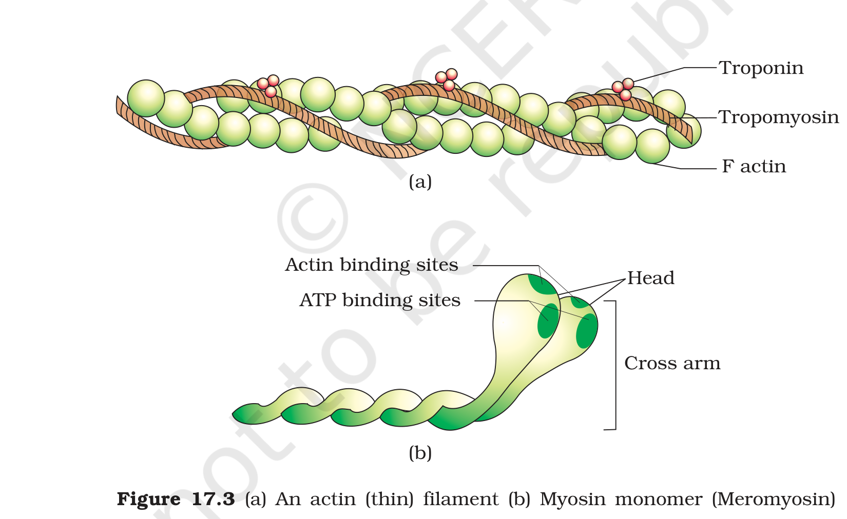
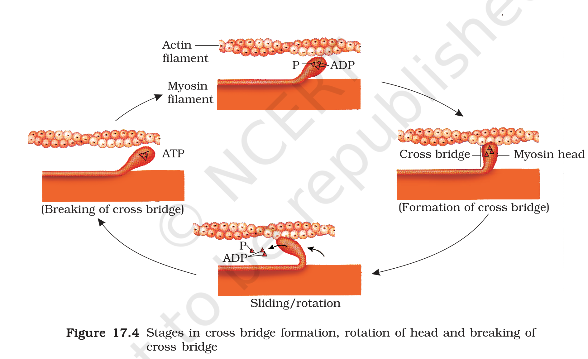
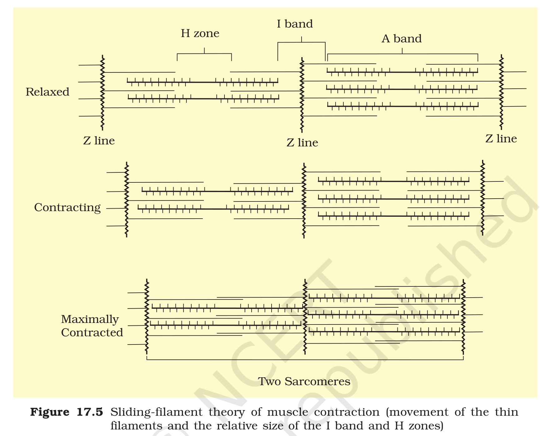
Figure 17.3: (a) an actin thin filament with troponin and tropomyosin and (b) a myosin monomer showing the head, cross arm and binding sites
Exam
The sliding filament answer must contain four links in order: calcium released from the sarcoplasmic reticulum, calcium binds troponin and tropomyosin moves, myosin head forms a cross bridge with actin, and the head tilts and pulls actin while ATP breaks the bridge. Finish with the changes in the bands: A band same, I band and H zone shorter, sarcomere shorter. The question on sarcomere bands is answered best with a quick labelled sketch.
Common mistake
Do not say that the filaments themselves shorten, because the filaments slide past one another. Do not say that calcium binds to myosin, because it binds to troponin. Do not describe the cross bridge as permanent, since ATP is required to break it. Do not forget that relaxation also needs energy, because the calcium must be pumped back into the sarcoplasmic reticulum.