

Concepts: Types of movement and the muscles that produce them
Locomotion and Movement24 min readLesson 1 of 7
1.1 Movement and locomotion
Living cells show movement of many kinds. A cell can push out a pseudopodium and crawl forward, the way a white blood cell or a macrophage does, and that is called amoeboid movement. Cilia on the surface of a cell beat in a coordinated wave and move the cell or the fluid around it, which is ciliary movement, seen in the ciliated epithelium of the respiratory tract and in the movement of the egg through the fallopian tube. A sperm cell swims with a single long flagellum, and that is flagellar movement. In a large animal all these cellular movements are not enough to move the whole body, so a special tissue, muscle, takes over, and the movement of the limbs and the body is called muscular movement.
Locomotion is the movement that carries the organism from one place to another. It needs more than muscle: the muscles pull on the bones, the joints decide the direction of the pull, and the nervous system decides when and how hard to pull. A running animal is therefore using the muscular, skeletal and neural systems together, and the support of the skeleton against gravity is what makes such movement possible.
1.2 The three types of muscle
Muscle tissue is of mesodermal origin. It is built of long cells which are called muscle fibres and which shorten when they are stimulated. The three types of muscle differ in structure and in control.
Skeletal muscle is attached to the bones and is the muscle that produces the movements of the body. Its fibres are long, cylindrical and striped, so it is called striated muscle. Its activity is under the conscious control of the brain through somatic nerves, so it is voluntary. The protein filaments inside it are arranged so regularly that the fibre looks striped under the microscope.
Smooth muscle is found in the walls of the gut, the blood vessels, the urinary bladder and other internal organs. Its cells are spindle shaped and have no stripes, so it is non-striated, and it works without conscious control, so it is involuntary. It moves food along the gut and squeezes the vessels, all without any decision from the conscious mind.
Cardiac muscle is the muscle of the heart wall. It is striated like skeletal muscle, but it is involuntary and it contracts rhythmically without rest. Its cells are joined end to end at special junctions and the tissue has its own rhythm, which is why the heart keeps beating even when a person is asleep. A fourth, related point is that the three types regenerate differently, but the classification asked in the examination is based on striations and on conscious control.
1.3 Inside a skeletal muscle
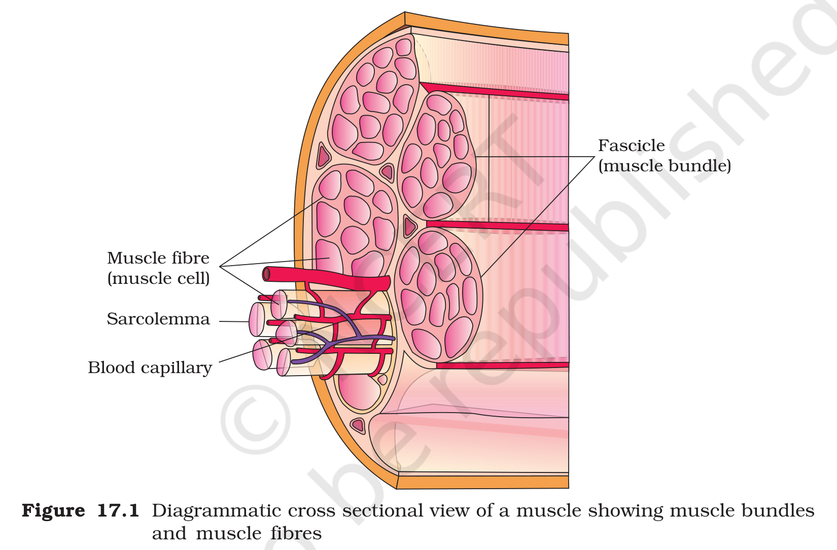
A skeletal muscle is made of many bundles of fibres, and the connective tissue around them carries the blood vessels and nerves. Each bundle is a fasciculus, each fibre inside the bundle is a muscle fibre, and each fibre contains many parallel threads called myofibrils. The thin plasma membrane of the fibre is the sarcolemma and its cytoplasm is the sarcoplasm. The sarcoplasm contains a large amount of endoplasmic reticulum, called the sarcoplasmic reticulum, and that membrane network stores calcium ions. A muscle fibre also contains many mitochondria because contraction uses a great deal of energy, and it stores the oxygen binding pigment myoglobin, which gives the red colour to some fibres.
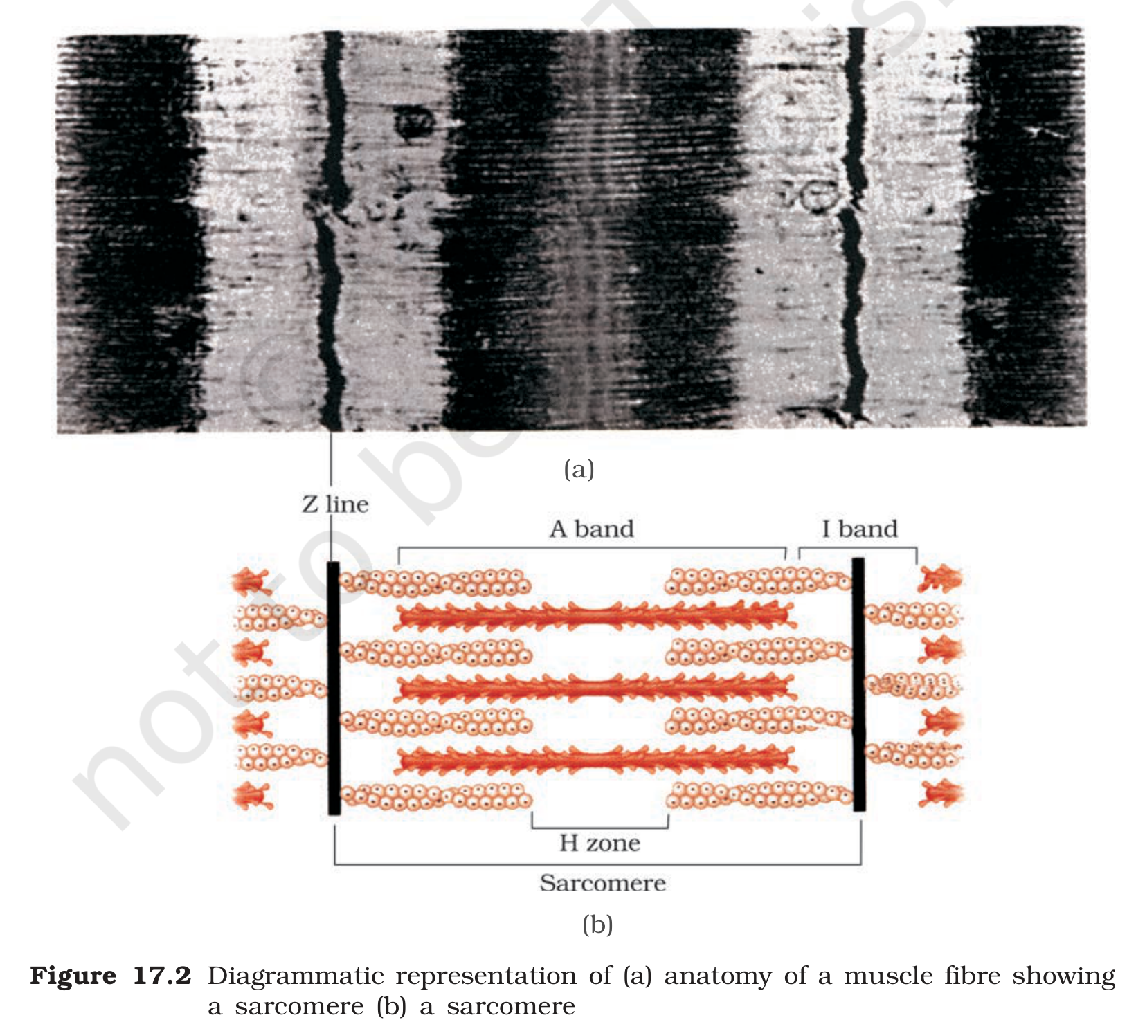
The myofibril carries alternating light and dark stripes. Myosin makes the dark stripe, which is named the A band, and actin makes the light stripe, the I band. Filaments of both kinds run parallel and can slide across one another. A paler region, the H zone, sits inside the A band, and a line through the middle of the H zone is the M line. The Z line crosses the middle of each I band, and the stretch of myofibril from one Z line to the next is a sarcomere. This is the unit whose shortening shortens the whole muscle.
Muscle fibres differ in their content of myoglobin. Red fibres, also called red muscles, are rich in myoglobin and in mitochondria, so they look dark red and can keep working for a long time. White fibres have less myoglobin and fewer mitochondria, so they look pale and tire sooner, but they can contract faster and more forcefully for short bursts. Most human muscles contain a mixture of the two fibre types, and the mixture decides whether a muscle is suited to endurance work or to a quick effort.
1.3.1 Three systems, one movement
A single step looks simple, but it needs three systems to act together. The muscles supply the force, the bones and joints form the levers that direct the force, and the nervous system decides which muscles to switch on and when. If the nerve supply to a muscle is cut, the muscle can no longer be ordered to contract, and if a joint is injured the strongest muscle cannot move the limb properly. This is why the chapter studies movement as a combined property of the muscular, skeletal and neural systems rather than as a property of muscle alone.
Exam
For the movement answer, give one example with each type: amoeboid for macrophages and leucocytes, ciliary for the respiratory epithelium, flagellar for the sperm, and muscular for the limbs. For the muscle types, draw a three row table with striations, control and one location for skeletal, smooth and cardiac muscle. Inside the muscle, the order from large to small is muscle, bundle, fibre, myofibril, sarcomere, filament.
Common mistake
Do not call cardiac muscle voluntary: it is striated but involuntary. Do not say smooth muscle is striated, because it has no stripes. Do not confuse the sarcolemma with the sarcoplasmic reticulum, since the sarcolemma is the membrane around the fibre and the sarcoplasmic reticulum is the calcium storing network inside it. Also do not say that the A band shortens when a muscle contracts; the width of the A band stays nearly the same while the I band and the H zone get smaller.