

Concepts: The pistil, the ovule and the embryo sac
Sexual Reproduction in Flowering Plants16 min readLesson 2 of 8
2.1 The pistil
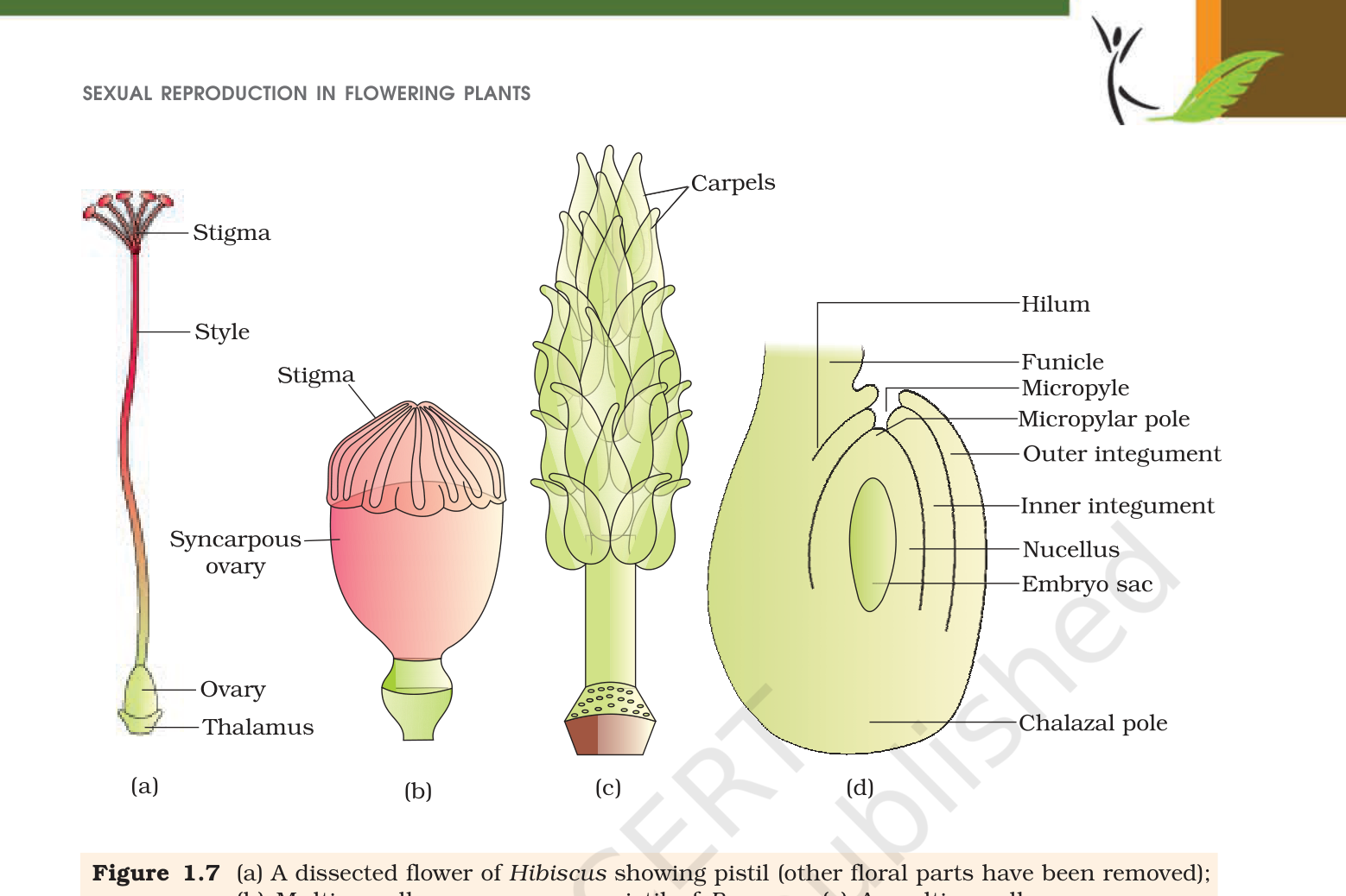
The gynoecium is the female part of the flower. It may have one pistil, which is the monocarpellary condition, or several pistils, which is multicarpellary. When there are several, they may be free or fused. Fused pistils are syncarpous, as in poppy, and free pistils are apocarpous, as in Michelia. Each individual pistil has three zones: the stigma at the top, the style in the middle and the ovary at the base.
The stigma is the landing platform for pollen. Its surface is often sticky or feathery, and the style lifts it above the ovary where insect visitors and air currents can reach it. The ovary is the swollen base and holds a cavity called the locule. Inside the locule sits the placenta, and the pattern of attachment of ovules to the placenta is what you studied in Class 11 as placentation. Each pistil can carry ovules of very different number. Wheat, paddy and mango have one ovule in the ovary, while papaya, watermelon and orchids carry many.
2.2 The ovule
An ovule attaches to the placenta by a short stalk, the funicle. The point where the body of the ovule merges with the funicle is the hilum. One or two protective layers, the integuments, wrap around the ovule and leave a small opening at the top called the micropyle. At the opposite end, where the integuments meet the base, is the chalaza. Inside the integuments lies the nucellus, a mass of cells full of reserve food, and inside the nucellus sits the embryo sac, the female gametophyte. A typical ovule has a single embryo sac.
The common textbook ovule is anatropous. It is bent so that the micropyle comes to lie close to the funicle, which places the micropylar end of the embryo sac next to the entrance the pollen tube will use. When you draw the ovule, mark the funicle and hilum on the outer side, the micropyle at the tip, the chalaza at the base and the nucellus inside. The pollen tube always enters through the micropyle.
2.3 Megasporogenesis
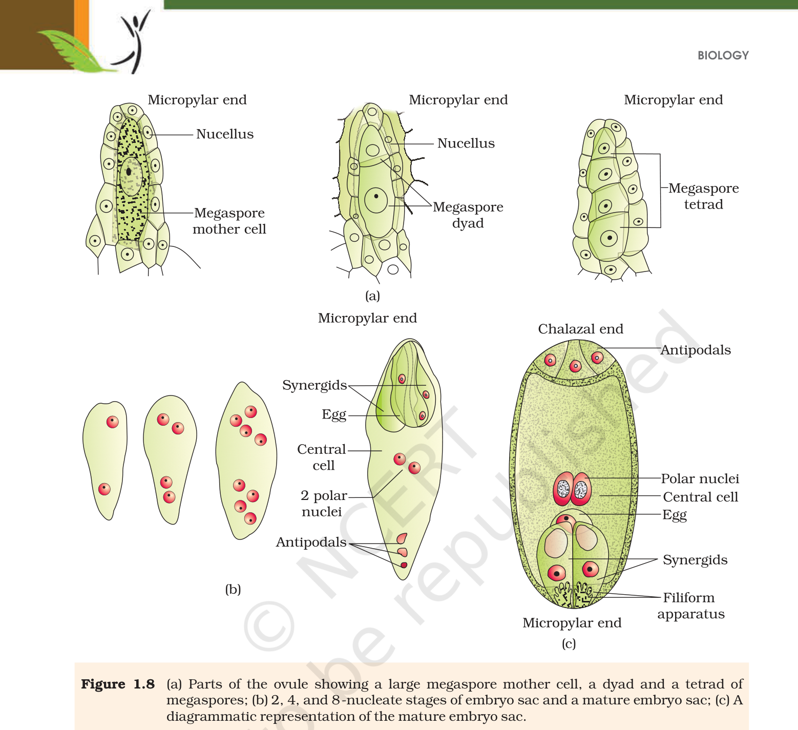
One cell of the nucellus near the micropylar end grows large, with dense cytoplasm and a prominent nucleus, and becomes the megaspore mother cell. Meiosis in this cell produces four haploid megaspores. In most flowering plants only one of the four survives. The other three degenerate, and the surviving megaspore becomes the embryo sac. Because the whole embryo sac comes from one megaspore, the process is called monosporic development.
The ploidy question that follows is a common exam item. The nucellus and the megaspore mother cell are diploid, both belonging to the sporophyte. The functional megaspore is haploid because it is a product of meiosis. The embryo sac that develops from it is also haploid in its nuclei and cells, except at the centre where two polar nuclei share one large cell.
2.4 Microsporogenesis and megasporogenesis side by side
The two processes look similar and boards use that confusion. Both start with a diploid mother cell, both use meiosis, and both end in a haploid product. The location and the fate of the four products differ.
| Feature | Microsporogenesis | Megasporogenesis |
|---|---|---|
| Site | Microsporangium of the anther | Nucellus of the ovule |
| Mother cell | Pollen mother cell | Megaspore mother cell |
| Division | Meiosis | Meiosis |
| Immediate product | A tetrad of four microspores | A tetrad of four megaspores |
| Survivors | All four become pollen grains | Usually only one survives |
| Final structure | Pollen grains | Embryo sac |
The survival difference is the part students skip. In the microsporangium the tetrad breaks apart and every microspore continues. In the nucellus three megaspores degenerate, and the single functional megaspore alone builds the female gametophyte. That is what the word monosporic records.
2.5 Numbers a good answer should carry
An embryo sac holds one egg. An ovule usually holds one embryo sac, formed from one functional megaspore. The number of ovules in an ovary then decides the possible seed number: one ovule can give one seed, as in wheat and mango, while papaya and watermelon carry many ovules and therefore many seeds. One ovary belongs to a pistil, and one flower can hold one pistil or many pistils. The chapter uses this counting chain at the end to remind you how large the reproductive output of a single tree can become.
2.6 From megaspore to embryo sac
The nucleus of the functional megaspore divides by mitosis. The two nuclei move to opposite ends and the sac is at the two-nucleate stage. Two more rounds of mitotic division give the four-nucleate and then the eight-nucleate sac. All three rounds are free nuclear, meaning the nuclei multiply without cell walls forming between them. Walls are laid down only after the eight-nucleate stage.
Look at how the eight nuclei are used. Three nuclei gather at the micropylar end and form the egg apparatus, which is two synergids plus one egg cell. The synergids have thickenings at their micropylar tip called the filiform apparatus, and this structure guides the pollen tube into the synergid. Three nuclei at the opposite, chalazal, end become the antipodal cells. The two remaining nuclei, the polar nuclei, stay in the middle of the large central cell.
Put together, the mature angiosperm embryo sac has eight nuclei but seven cells: two synergids, one egg, three antipodals and one central cell holding two polar nuclei. That sentence is worth memorising in both directions, because questions ask for the cell count and the nuclei count separately.
| Structure | Ploidy | Point to remember |
|---|---|---|
| Nucellus | 2n | Provides the megaspore mother cell |
| Megaspore mother cell | 2n | Undergoes meiosis |
| Functional megaspore | n | Only one of four survives |
| Embryo sac cells | n | Seven cells, eight nuclei |
| Central cell | n + n polar nuclei | Becomes triploid after fusion with a male gamete |
Exam
A three-mark diagram question usually asks for a labelled embryo sac. Label the micropylar end with synergids and egg, the chalazal end with antipodals, and the middle with the two polar nuclei. Write both numbers, eight nuclei and seven cells, and show where syngamy and triple fusion will happen.
Common mistake
Do not write that all four megaspores form embryo sacs. Only one does, and the rest degenerate. Do not call the synergids eggs; they are helper cells that guide the tube, and the egg is the single cell between them. The filiform apparatus belongs to the synergids, not to the egg.