

Seeing the invisible world
The Invisible Living World13 min readLesson 1 of 5
1.1 A world hidden by size
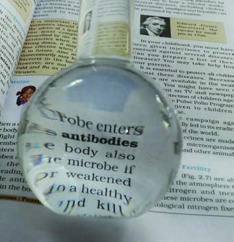
The human eye can see only objects above a certain size. For a very long time, the tiny living things around us stayed unknown simply because they were too small to notice. A curved piece of glass changed that. The glass was thicker at the centre and thinner at the rim, rather like a lentil seed, and the name lens comes from that shape. Reading glasses, magnifying glasses and microscopes all use lenses, and each improvement helped people look further into the small world.
1.2 A flask of water as a magnifier
You can test the idea with a simple set-up. Fill a round-bottom flask with water, close its mouth with a cork, and place it on an open book. The letters seen through the flask look larger. The water-filled glass behaves like a magnifying glass. A real magnifying glass shows the fine details of an ant's body, details that the unaided eye misses.
Tools do not replace our senses; they extend them. A ruler turns "long" into centimetres, and a lens turns "too small to see" into "visible". Each new instrument gives the investigator a sharper look at the world.
1.3 Hooke finds empty rooms in cork
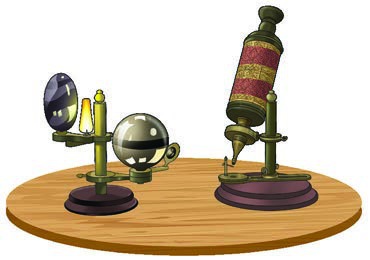
In 1665 Robert Hooke brought out a book titled Micrographia. He watched specimens closely and drew them with skill, and the book showed detailed pictures of tiny objects that people had never seen before. His microscope made things look about 200 to 300 times larger than the unaided eye could manage, which was a great advance for that time.
One day Hooke looked at a thin slice of cork. He found it full of tiny empty compartments, packed together somewhat like a honeycomb. Each little compartment was drawn and named a cell, and that use of the word cell for the basic unit of life was new in science. The name has stayed ever since.
1.4 Leeuwenhoek opens the microbial world
Around the same period, in the 1660s, the Dutch scientist Antonie van Leeuwenhoek made better lenses and built more useful microscopes. He became the first to describe living things as small as bacteria and blood cells after seeing them clearly. Because of that work, he is remembered as the Father of Microbiology.
The two discoveries fit together. Hooke showed that living material is built of small units, and Leeuwenhoek showed that some of those units live on their own as complete organisms.
1.5 Why a curved glass magnifies
A lens works because light bends when it passes from one material into another. A curved piece of glass, or a flask full of water, changes the direction of light rays and makes them spread out or come together in a way that creates a larger image. The eye receives that larger image, so a small object appears bigger. The same principle sits inside every microscope, where a combination of lenses produces a much larger, clearer image than a single hand lens can.
The idea is easy to try at home with a drop of water on a leaf or a glass of water held above a page. The curved surface does the work, and the object looks changed in size. Once people understood this effect, improving the shape and quality of lenses became a practical goal.
1.6 From curiosity to instrument
Hooke and Leeuwenhoek were investigators in the full sense. They built or improved instruments, looked patiently at specimens, drew what they saw and shared their findings with other people. Their drawings were part of the evidence, because a careful sketch could be checked by another observer.
Microscopes improved over the following centuries. The basic parts of a school microscope are a lens system, a stage for the slide, a light source and a focusing arrangement. Each part solves one problem: the lens magnifies, the stage holds the specimen steady, the light makes the details visible, and the focus brings the image into clear view.
1.7 What counts as living
Size does not decide whether something is alive. A microbe that is a single cell can still grow, take in food, release waste, respond to its surroundings and reproduce. Its small size only means that we need a tool to see it. The chapter therefore moves from the instruments to the cells they reveal, and then to the organisms that exist as single cells or as tiny clusters of them.
1.8 Better tools, deeper views
The microscopes used in school laboratories magnify a specimen about 100 to 400 times. A foldable paper microscope costs much less and can be carried anywhere; it does not give the same detail as a laboratory instrument, but it makes the tiny world available to many more people. For looking at the structures inside a cell, scientists use an electron microscope, which can magnify about 10,00,000 times. This chapter uses these tools to visit two hidden worlds: the cells that build living bodies, and the microorganisms that live all around us.
1.9 Handling a microscope
A microscope is a sturdy instrument, but its lenses are delicate. Carry it with both hands, keep the lenses clean with soft tissue, and lower the objective slowly while watching from the side so the lens does not touch the slide. Begin with the lowest magnification, find the specimen, and then change to a higher lens. A scratch on a lens can spoil every later observation, so careful handling is part of good method.