

The many kinds of microorganisms
The Invisible Living World13 min readLesson 3 of 5
3.1 Living things too small to see
Some living organisms are made of just one or a very few cells, and they are far too small to be seen without a microscope. These are microorganisms, or microbes. The word comes from micro, meaning very small, and organisms, meaning living beings. Microbes live in water, in soil, in air, and even inside the bodies of plants and animals, including us.
Some microbes are single-celled, or unicellular, such as bacteria and Amoeba. Others are multicellular, such as some fungi and algae. Yeast has one cell, whereas moulds have many. To see their cells, we again need a microscope, which enlarges them and makes them visible.
3.2 Sampling a pond and a handful of soil
Collect a little pond water or water from a stagnant puddle with the help of an adult. Place a drop on a slide, add a coverslip and observe under a microscope. In pond water you may find Amoeba, a single cell with an irregular shape that moves about; Paramecium, also a single cell, which moves with the help of specialised structures; and algae, single-celled organisms that look green because of the green pigment chlorophyll.
A soil suspension reveals another set of organisms. Mix some moist soil with water, let the mixture settle, and take a drop from the top layer. You may find moulds with branched filaments and no chlorophyll, algae with spherical green cells, and bacteria in several shapes. Bacteria may be spherical, comma-shaped, spiral or rod-shaped, and many have hair-like projections that help them move.
3.3 The groups at a glance
Observations of pond water and soil give the same categories that scientists use to group microbes.
| Group | What it is like | Examples |
|---|---|---|
| Bacteria | Unicellular; no well-defined nucleus, a nucleoid instead; several shapes | Rods, spheres, spirals |
| Protozoa | Unicellular animals that move about | Amoeba, Paramecium |
| Fungi | Cell wall, no chlorophyll; yeast is unicellular, moulds are multicellular | Yeast, bread moulds |
| Algae | Plant-like, contain chlorophyll and make their own food; many are microscopic | Microalgae such as Spirulina |
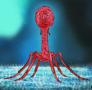
| Viruses | Acellular, not a cell at all; multiply only inside a living host cell | Bacteriophage, disease viruses |
The table shows how varied the invisible world is. It also shows why the earlier discovery mattered: without a microscope, none of these organisms would be visible at all, and the microscope made them available for careful study.
3.4 Everywhere, even in the extremes
Microbes turn up in surprising places. They live in hot water springs, in snow-cold zones and in ordinary moderate climates. They grow on the surfaces of leaves, stems and roots. Some find a home inside the human gut, where they help with digestion. Their shapes, sizes and structures are as diverse as their addresses.
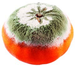
Food left outside for a few days may develop a powdery or cotton-like covering, a sign that microorganisms are using the food as a source of nutrients. The orange in the figure has been infected by a mould, a kind of fungus.
Microscopes for this work magnify about 100 to 400 times. A low-cost foldable paper microscope may not show the same detail as a laboratory instrument, but it is cheap and portable, which brings a view of this hidden world to many more classrooms and communities.
Viruses need a special mention. They are microscopic, but they are not cells: they are acellular and can multiply only after entering a living host cell. They may infect plants, animals or bacteria, and they often cause disease. That is why viruses are usually described separately from bacteria, protozoa, fungi and algae rather than as one more simple group of cells.
3.5 How the tiny ones move
Movement is one of the easiest things to notice in a drop of pond water. Amoeba creeps along by changing the shape of its body, which is why its outline keeps shifting. Paramecium moves with the help of specialised structures on its surface, and algae may also move with their own structures. Many bacteria have one long hair-like structure or many tiny hair-like projections around it, and these help them travel.
Movement is not the only sign of life. A microbe may be seen taking in material, growing longer before dividing, or forming spores. A single drop of water can therefore show a small community in action rather than a still picture.
3.6 Observing like a scientist
A useful observation starts with a clean slide, a small drop, a coverslip and a steady focus. Move the slide slowly across the field and look for differences in shape, colour and movement. Make a simple drawing for each kind of organism, note its colour and movement, and write down the magnification used. Taking a second drop from the same sample helps check whether the first observation was typical.
Identification begins with grouping. Round green cells with chlorophyll look like algae, irregular moving cells like Amoeba, slipper-shaped moving cells like Paramecium, and branched filaments without chlorophyll like moulds. Bacterial cells come in several shapes and need a stronger lens to show their details clearly.
3.7 From observation to classification
Scientists group microorganisms by features that can be observed: whether the organism has one cell or many, whether it has a well-defined nucleus, whether it contains chlorophyll, and what shape its cells take. These features produce the groups described in the table. A single drop from a pond is only a sample of a much larger community, so repeated observations from different places give a better picture of microbial diversity.