

The cell: the basic unit of life
The Invisible Living World15 min readLesson 2 of 5
2.1 The building blocks of living things
All living organisms are made of cells. A cell is the basic unit of life, and the body of every plant and animal is built from many of them. Some living things, such as bacteria and Amoeba, manage with just one cell, while others are made of millions. In an organism with many cells, each cell carries out its own jobs and also cooperates with the rest.
2.2 Two slides: onion peel and cheek cells
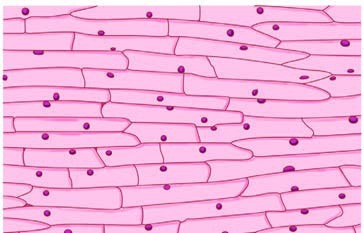
You can see plant cells with a simple onion peel. The thin transparent layer from the inner surface of an onion is stained with safranin, rinsed, mounted in glycerin, and covered with a coverslip. The stain gives the cells a pink colour and improves contrast; the glycerin keeps them from drying out. Under the microscope, the onion peel shows nearly rectangular cells packed closely together with no gaps between them. A wall made of bricks is a useful picture of how such cells are arranged.
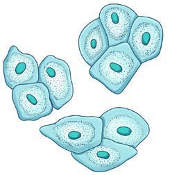
Animal cells can be seen in a cheek scrape. The inside of the cheek is gently scraped with the blunt end of a clean toothpick, the material is spread on a slide and stained with methylene blue, and a coverslip is added. Under the microscope the cheek cells look polygon-shaped and thin. Staining helps because it increases contrast, and the cells become much easier to see.
2.3 The three basic parts
Most cells share three main parts. The cell membrane is the outer boundary. It encloses the cell, separates it from its neighbours, and is porous, letting in materials needed for life and letting out waste. Inside the membrane is the cytoplasm, a jelly-like material that holds the other cell components and compounds including carbohydrates, fats, proteins and mineral salts. Most life processes take place in the cytoplasm. The nucleus, a round structure with its own thin membrane, regulates the activities of the cell and its growth.
Plant cells have an extra layer outside the cell membrane, called the cell wall. The wall gives rigidity and strength, which is why plant cells sit compactly together and give the plant firm support.
2.4 Extra parts in plant cells
Plant cells also contain small rod-shaped structures called plastids. Some plastids, such as chloroplasts, contain chlorophyll, the green pigment that carries out photosynthesis. In non-green parts of a plant, plastids help store substances. Plant cells also contain a big space known as a vacuole, which stores useful substances, removes waste and helps the cell keep its shape. Animal cells usually have small vacuoles or none at all.
| Feature | Plant cell | Animal cell |
|---|---|---|
| Cell wall | Present | Absent |
| Chloroplast | Usually present | Absent |
| Large vacuole | Present | Small vacuoles or none |
| Shape | Often rectangular and fixed | Often rounded and variable |
| Nucleus | Present | Present |
Bacterial cells are different again. They have no well-defined nucleus and no nuclear membrane; instead the genetic material sits in a region called the nucleoid. Fungal cells have a cell wall but no chloroplasts, so making their own food by photosynthesis is impossible for them.
2.5 One shape, one job
Cells vary in shape and structure, and the shape usually fits the job. A muscle cell is spindle-shaped, which helps it contract and relax. A nerve cell is long and branched, which helps it carry messages quickly to different parts of the body. Cheek cells are thin and flat, which suits their role as a protective lining. In plants, cells may be rectangular, elongated, oval or tube-like, and some form long tubes that carry water.
The digestive tract shows how cells cooperate. Muscle cells in the food pipe contract and relax in a wave-like rhythm that pushes food towards the stomach. Muscle cells in the stomach wall churn the food, while other cells in the lining produce digestive juices and acid. No single cell does all the work; together they make digestion possible.
2.6 From cell to organism
A group of similar cells forms a tissue. Different tissues together form an organ, several organs work together as an organ system, and all the organ systems make one organism. The order is cell, tissue, organ, organ system, organism, and a brick wall, a room and a building follow the same idea.
A single cell can also start a whole multicellular organism. The yolk of an ostrich egg is one cell, and at about 130 to 170 millimetres across it is the largest known cell. The egg divides repeatedly and builds a complete living being. The life of a complex organism begins with that one cell.
2.7 Preparing a slide
Both cell activities follow the same practical pattern. A very thin specimen is taken, because thick material scatters light and hides details. It is stained to increase contrast, mounted in glycerin to stop it drying, and covered with a coverslip so the material stays flat and the lens stays clean. Air bubbles are avoided because they look like dark circles and confuse the observer.
The slide is then placed on the stage, the light is adjusted, and the focus is moved slowly until the cells appear. Drawing what is seen, rather than what is expected, is part of the habit of an investigator. A drawing with labels becomes a record that can be compared with another preparation, or with a textbook figure.
2.8 Cells in the lab and in the body
A first look through the microscope shows that cells are not identical. Onion peel cells are box-like and tightly packed; cheek cells are flat and irregular; a nerve cell has long branches; a muscle cell is spindle-shaped. A single drop from a culture or a scrape from the cheek can hold thousands of cells, and most of them are far too small to see without magnification.
The ostrich egg is the exception that proves the rule. It is a single cell large enough to hold in the hand, but most cells are microscopic. What matters is not the size of the cell but the fact that every living thing, from a bacterium to a tree to a human being, is built from one or more of these units.